组成图示
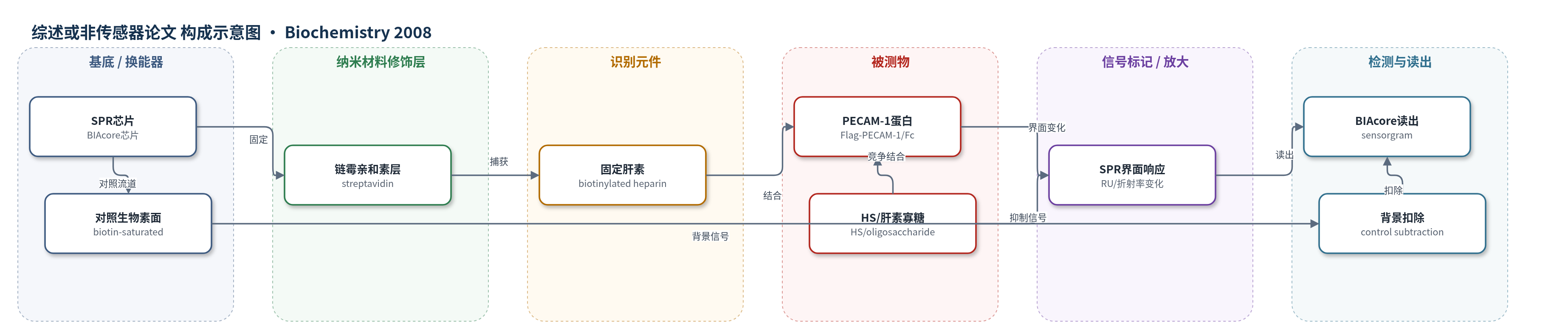
点击图片查看大图 · 依据论文自动绘制
传感器类型
综述或非传感器论文
检测对象
血小板内皮细胞黏附分子1(PECAM-1/CD31)、肝素(heparin)、硫酸乙酰肝素(HS);样品基质:纯化蛋白溶液、SPR固定相、A2058黑色素瘤细胞表面
检测原理
本研究以SPR芯片为换能器,将还原端生物素化肝素通过链霉亲和素固定于芯片表面。在pH 6.3的Bis-Tris/NaCl缓冲液中,流动相注入Flag-PECAM-1或PECAM-1-Fc。PECAM-1外源域中域2/3区域与肝素发生特异性结合,结合位点涉及域2的K176、L177、R179及域3的H239、K255、H253等碱性/组氨酸残基。蛋白结合使芯片界面质量/折射率增加,SPR共振信号以响应单位(RU)升高;RU随PECAM-1浓度增加而增加,解离后信号下降。结合对pH敏感,pH 6.3时组氨酸质子化有利于结合,pH 7.4明显减弱。肝素/HS或特定肝素寡糖可与PECAM-1预混竞争,降低RU,从而反映结合特异性。无酶或纳米放大。
检测灵敏度
原文未报告LOD、线性范围、灵敏度斜率或相关系数。
效应效果
两种表达系统均显示pH 6.3结合固定肝素,pH 7.4明显减弱。域删除实验表明域1-Fc不结合,域1-2-Fc弱,域1-3-Fc及全长良好;ELISA支持域2/3为主位点、域5/6次要。肝素和HS有效抑制结合,硫酸软骨素C/A几乎不抑制,硫酸软骨素B高浓度仅轻微抑制。氯酸盐处理使A2058细胞HepSS-1信号降低81%,Flag-PECAM-1结合降至阴性对照;heparinase III处理也显著降低结合。Flag-D1-3细胞信号约阴性对照5倍。DP12 HS片段与全长HS同样有效抑制SPR结合。作者认为其可调节酸性微环境中白细胞外渗和肿瘤转移。
传感器的构成
- 基底/换能器:BIAcore 2000 SPR芯片(streptavidin sensor chip),提供表面等离子共振换能表面
- 修饰层:链霉亲和素(streptavidin)表面,用于高亲和捕获生物素化肝素
- 识别元件:还原端生物素化肝素(biotinylated heparin),固定于链霉亲和素表面,作为PECAM-1配体
- 对照表面:第二流道饱和生物素(biotin)表面,用于扣除非特异背景
- 流动相样品:Flag-PECAM-1或PECAM-1-Fc外源域蛋白,溶于10 mM Bis-Tris、150 mM NaCl、0.005% Tween-20、2 mM EDTA,pH 6.3
- 竞争/抑制元件:肝素、硫酸乙酰肝素(HS)或肝素寡糖,与PECAM-1预混以竞争结合
- 信号读出:SPR响应单位(RU)随界面结合量变化,由BIAcore 2000记录传感器图
中文摘要
血小板内皮细胞黏附分子1(PECAM-1,CD31)是免疫球蛋白超家族细胞黏附分子,含六个Ig样结构域,参与炎症时白细胞外渗和血管内皮完整性维持。尽管已知PECAM-1通过结构域1同型结合介导细胞黏附,但已有多种异型配体被提出。本研究重新考察肝素和硫酸乙酰肝素(HS)是否为PECAM-1配体。PECAM-1外源域先以与人IgG1 Fc区融合的结构域6融合蛋白表达,再以结构域1 N端Flag标签表达(Flag-PECAM-1)。两种蛋白在表面等离子共振(SPR)结合实验中均结合固定于生物传感器芯片上的肝素。结合具有pH敏感性,但在略酸性pH下易于测量。对两种表达系统中制备的一系列PECAM-1结构域缺失体进行肝素结合检测,发现主要肝素结合位点需要结构域2和3。Flag-PECAM-1及含结构域1-3的Flag蛋白结合黑色素瘤细胞表面HS,而含结构域1-2的Flag蛋白不结合。肝素寡糖可抑制Flag-PECAM-1结合固定肝素,某些结构抑制活性更强。分子建模同样确定结构域2/3交界为肝素结合位点,并揭示艾杜糖醛酸构象对结合的重要性。PECAM-1确实结合肝素/HS,但其位点与同型结合所需位点不同。
英文摘要
Platelet endothelial cell adhesion molecule 1 (PECAM-1) (CD31), a member of the immunoglobulin (Ig) superfamily of cell adhesion molecules with six Ig-like domains, has a range of functions, notably its contributions to leukocyte extravasation during inflammation and in maintaining vascular endothelial integrity. Although PECAM-1 is known to mediate cell adhesion by homophilic binding via domain 1, a number of PECAM-1 heterophilic ligands have been proposed. Here, the possibility that heparin and heparan sulfate (HS) are ligands for PECAM-1 was reinvestigated. The extracellular domain of PECAM-1 was expressed first as a fusion protein with the Fc region of human IgG1 fused to domain 6 and second with an N-terminal Flag tag on domain 1 (Flag-PECAM-1). Both proteins bound heparin immobilized on a biosensor chip in surface plasmon resonance (SPR) binding experiments. Binding was pH-sensitive but is easily measured at slightly acidic pH. A series of PECAM-1 domain deletions, prepared in both expression systems, were tested for heparin binding. This revealed that the main heparin-binding site required both domains 2 and 3. Flag-PECAM-1 and a Flag protein containing domains 1-3 bound HS on melanoma cell surfaces, but a Flag protein containing domains 1-2 did not. Heparin oligosaccharides inhibited Flag-PECAM-1 from binding immobilized heparin, with certain structures having greater inhibitory activity than others. Molecular modeling similarly identified the junction of domains 2 and 3 as the heparin-binding site and further revealed the importance of the iduronic acid conformation for binding. PECAM-1 does bind heparin/HS but by a site that is distinct from that required for homophilic binding.