组成图示
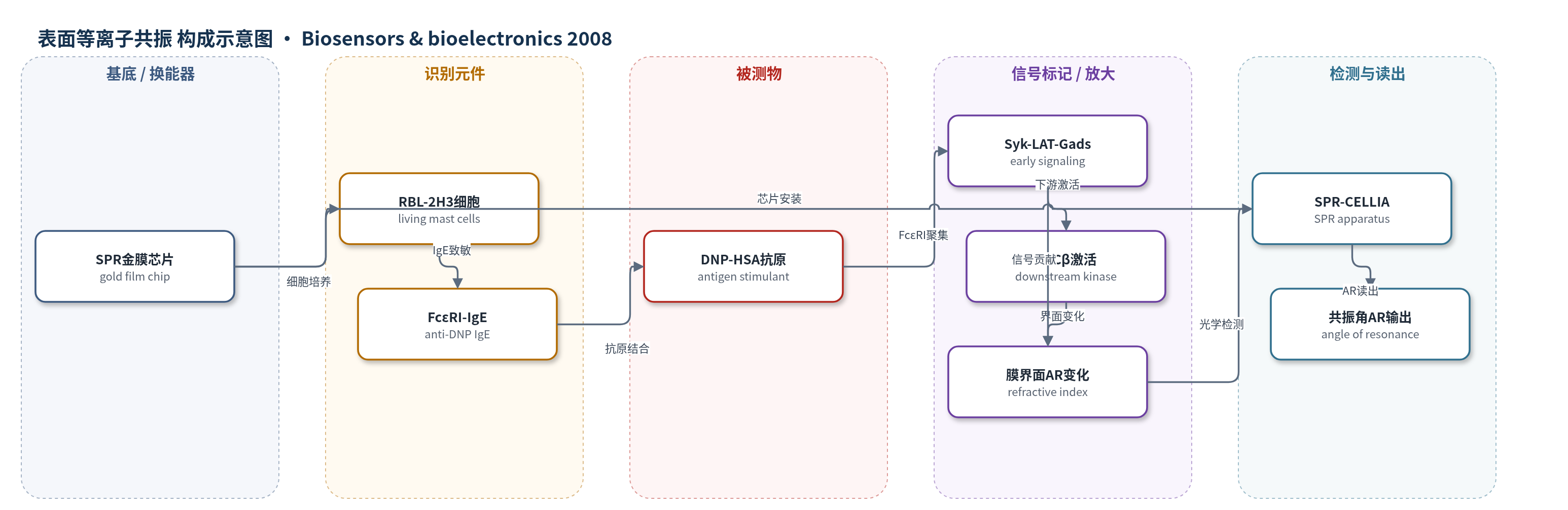
点击图片查看大图 · 依据论文自动绘制
传感器类型
表面等离子共振(SPR)生物传感器
检测对象
抗原刺激的RBL-2H3肥大细胞激活(DNP-HSA抗原;样品基质:葡萄糖生理盐水/Pipes缓冲液中的贴壁细胞)
检测原理
SPR生物传感器利用金膜表面产生的倏逝场,实时监测传感芯片附近约数百纳米范围内折射率或质量变化引起的共振角(AR)偏移。将RBL-2H3肥大细胞培养于芯片表面,并用抗DNP IgE致敏后,DNP-HSA抗原交联细胞表面FcεRI,启动Syk、LAT、Gads等早期信号事件,并进一步激活PKCβ。这些细胞内事件引起细胞膜及膜邻近区域的结构、电荷或折射率变化,使AR增大。信号无需外源标记,AR变化幅度反映FcεRI依赖的细胞激活程度;抑制Syk、LAT、Gads或PKCβ会显著降低AR变化,而Grb2/Ras-MAPK通路缺失不影响AR,说明SPR读出主要对应PKCβ下游的膜界面事件。
检测灵敏度
未报告LOD、线性范围、灵敏度斜率或相关系数。
效应效果
该SPR方法可实时、无标记地检测活肥大细胞抗原激活,且信号具有明确的通路特异性。过表达显性负性Syk或SLAP使抗原诱导AR仅轻微增加;显性负性LAT或Gads几乎完全抑制AR增加;显性负性Grb2虽抑制Ras-MAPK激活,但不影响AR。除PKCβ外,过表达PKCα、δ、ε、γ均降低AR增幅,PKCβ siRNA也降低AR,表明PKCβ对AR增加起正调控作用,其他PKC亚型呈负调控。PMA单独刺激仅产生较小SPR信号,提示FcεRI聚集与钙依赖PKC激活共同决定信号。文中数据为三次独立实验的代表性结果,未报告RSD、回收率或与ELISA/qPCR的定量对比。
传感器的构成
- 基底/换能器:金膜SPR传感芯片,提供倏逝场并产生共振角AR信号
- 识别元件:RBL-2H3肥大细胞,贴壁培养于芯片表面,作为活细胞识别与信号发生单元
- 识别元件:抗DNP IgE(SPE-7),致敏肥大细胞表面FcεRI,介导抗原特异性识别
- 刺激物:DNP-HSA抗原,交联FcεRI并触发细胞激活
- 缓冲液:葡萄糖生理盐水/Pipes缓冲液,维持细胞状态并控制SPR背景
- 读出仪器:SPR-CELLIA,实时监测共振角AR变化
中文摘要
表面等离子共振(SPR)生物传感器在将RBL-2H3肥大细胞培养于传感芯片并用抗原刺激时,可检测到共振角(AR)的显著变化,但导致这种大AR变化的分子事件尚不清楚。本研究通过遗传操纵细胞内信号分子,探讨抗原诱导的细胞内信号事件与AR变化之间的关系。过表达显性负性Syk或SLAP的RBL-2H3细胞在抗原刺激后AR仅轻微增加;过表达显性负性LAT或Gads则几乎完全抑制抗原诱导的AR增加。除PKCβ外,过表达其他PKC亚型均使抗原诱导的AR增幅降低;针对PKCβ的小干扰RNA也抑制了抗原诱导的AR增加。结果表明,Syk、LAT、Gads的激活以及随后的PKCβ激活是SPR生物传感器检测到的肥大细胞抗原诱导AR增加所必需的。
英文摘要
Surface plasmon resonance (SPR) biosensors detect large changes of angle of resonance (AR) when RBL-2H3 mast cells are cultured on a sensor chip and stimulated with antigen. However, the detail of molecular events that are responsible for such large changes of AR remained unknown. In this study, we investigated the relationship between intracellular signaling events induced by antigen and the change of AR, by genetic manipulation of intracellular signaling molecules; spleen tyrosine kinase (Syk), src-like adaptor protein (SLAP), linker for activation of T cells (LAT), growth-factor-receptor-bound protein 2 (Grb2), Grb2-related adaptor protein (Gads), and isotypes of protein kinase C (PKC). RBL-2H3 mast cells overexpressing dominant-negative Syk or SLAP, which both interfere with active Syk, exhibited only minimal increase of AR in response to antigen stimulation. Likewise, the interference of the activation of LAT and Gads, by expressing dominant-negative LAT and Gads, respectively, resulted in nearly complete suppression of the antigen-induced increase of AR. The cells overexpressing PKCs, apart from PKCbeta, showed a reduced extent of increase of AR in response to antigen stimulation. Moreover, the introduction of the small interfering RNA targeted against PKCbeta suppressed the antigen-induced increase of AR. These results indicate that the activation of Syk, LAT, Gads, and subsequent PKCbeta is indispensable for the antigen-induced increase of AR of mast cells detected by SPR biosensors.