组成图示
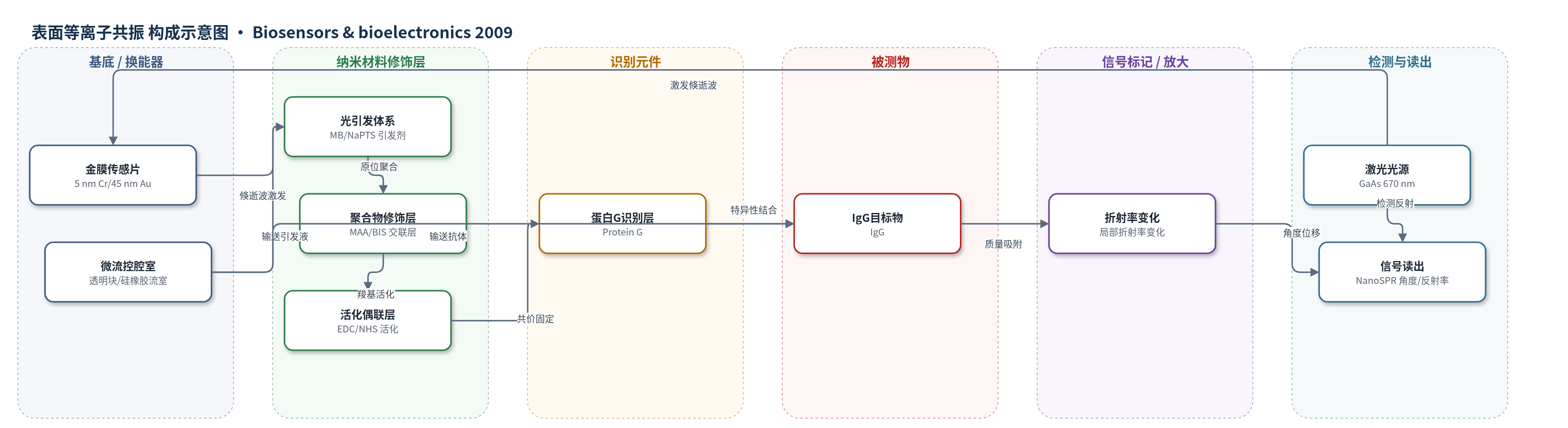
点击图片查看大图 · 依据论文自动绘制
传感器类型
表面等离子共振(SPR)生物传感器
检测对象
免疫球蛋白 G(IgG),样品基质为 10 mM 醋酸缓冲液(pH 5.0)
检测原理
本传感器基于表面等离子共振(SPR)直接检测界面质量/折射率变化。670 nm 激光经 ATR 棱镜在金膜表面激发倏逝波,MB/NaPTS 光氧化还原体系在该场中产生自由基,使 MAA 与 BIS 仅在探测点原位聚合为交联 poly(MAA) 层。EDC/NHS 活化聚合物羧基后共价固定 Protein G。当 IgG 流经表面并与 Protein G 特异性结合时,金膜附近局部折射率增加,SPR 共振角发生位移;结合量越大,角度/反射率变化越大。信号由 NanoSPR-321 实时读出,0.1 M HCl 可解离 IgG 实现再生。该体系未使用酶催化或核酸扩增,属于直接 SPR 质量传感。
检测灵敏度
原文未报告具体 LOD、线性范围、灵敏度斜率或相关系数。
效应效果
该免疫传感器以 Protein G–IgG 为模型体系,工作通道在 IgG 结合时出现明显 SPR 角位移,再生后响应可重复;参考通道无 Protein G,仅显示非特异吸附,说明识别层具有特异性。聚合物斑点可耐受洗涤及化学处理,蛋白 G 共价固定后在三个结合–解离–再生循环中响应一致,稳定性与重现性良好,原文未报告 RSD 或实际样品回收率。作者指出其性能与常规 Biacore 固定方法相当,并可在 2–3 h 内由裸金 SPR 芯片构建工作传感器。由于识别材料仅沉积于探测点,理论上可提高昂贵受体利用效率、降低非探测区结合损失,并有利于微型化与多传感阵列扩展。
传感器的构成
- 基底/换能器:玻璃棱镜(n=1.6160)与金膜传感片(5 nm Cr/45 nm Au),Au 层产生表面等离子体倏逝波
- 微流控腔室:透明块与约 1 mm 硅橡胶间隔件形成双通道流室,用于输送缓冲液、引发液和样品
- 光引发体系:亚甲基蓝(MB)与对甲苯磺酸钠(NaPTS)组成光氧化还原引发剂,在 670 nm 倏逝波场中产生自由基
- 聚合物修饰层:甲基丙烯酸(MAA)与 N,N-亚甲基双丙烯酰胺(BIS)原位聚合形成交联 poly(MAA) 层,提供羧基功能化支撑
- 偶联活化剂:EDC 与 NHS 活化 poly(MAA) 羧基生成 NHS 酯,用于共价固定蛋白 G
- 识别元件:蛋白 G(Protein G)共价固定于聚合物层,特异性结合 IgG
- 封闭剂:乙醇胺(ethanolamine)封闭未反应 NHS 酯,减少非特异结合
- 信号标记:无外源标记,IgG 结合引起局部折射率变化,由 SPR 角度/反射率变化直接检测
中文摘要
传统表面等离子共振(SPR)传感凝胶层常通过沉淀、旋涂或全表面原位聚合等整体方法制备,并将抗体或受体共价固定于凝胶表面。由于激光仅探测共振点,这种方式在材料利用上较为浪费。本文提出利用 SPR 器件表面的倏逝波场能量,在激光探测点原位形成聚合物层,从而提高抗体、酶、适配体、聚合物或分子印迹聚合物等识别元件的利用效率。研究采用亚甲基蓝(MB,敏化染料)与对甲苯磺酸钠(NaPTS,还原剂)组成光氧化还原引发剂体系,在 670 nm 激光激发下,于 SPR 焦点处聚合 N,N-亚甲基双丙烯酰胺(BIS)与甲基丙烯酸(MAA)水溶液;溶液及其他表面位置未发生聚合。通过改变单体浓度、染料浓度和曝光时间,可精确控制聚合物厚度(20–200 nm)。随后采用 EDC/NHS 标准羧基活化偶联法固定蛋白 G(Protein G),并以 IgG 结合为模型验证免疫传感器性能。结果表明该方法可制备功能化聚合物识别层,具有良好重复性,验证了沉积方法。
英文摘要
Traditionally, the integration of sensing gel layers in surface plasmon resonance (SPR) is achieved via "bulk" methods, such as precipitation, spin-coating or in-situ polymerization onto the total surface of the sensor chip, combined with covalent attachment of the antibody or receptor to the gel surface. This is wasteful in terms of materials as the sensing only occurs at the point of resonance interrogated by the laser. By isolating the sensing materials (antibodies, enzymes, aptamers, polymers, MIPs, etc.) to this exact spot a more efficient use of these recognition elements will be achieved. Here we present a method for the in-situ formation of polymers, using the energy of the evanescent wave field on the surface of an SPR device, specifically localized at the point of interrogation. Using the photo-initiator couple of methylene blue (sensitizing dye) and sodium p-toluenesulfinate (reducing agent) we polymerized a mixture of N,N-methylene-bis-acrylamide and methacrylic acid in water at the focal point of SPR. No polymerization was seen in solution or at any other sites on the sensor surface. Varying parameters such as monomer concentration and exposure time allowed precise control over the polymer thickness (from 20-200 nm). Standard coupling with 1-ethyl-3-(3-dimethylaminopropyl)carbodiimide and N-hydroxysuccinimide was used for the immobilization of protein G which was used to bind IgG in a typical biosensor format. This model system demonstrated the characteristic performance for this type of immunosensor, validating our deposition method.