组成图示
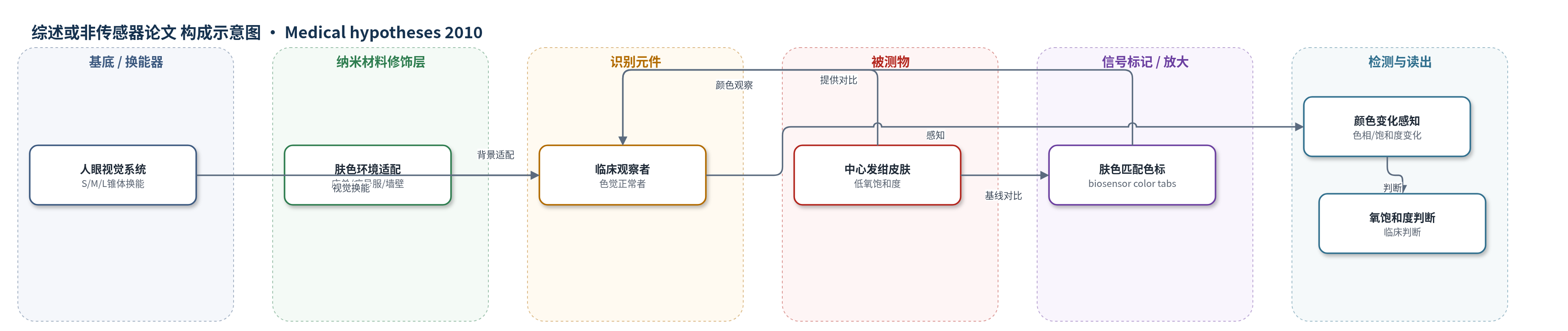
点击图片查看大图 · 依据论文自动绘制
传感器类型
综述或非传感器论文
检测对象
中心发绀(central cyanosis)、动脉氧饱和度(arterial oxygen saturation);样品基质:皮肤、唇、舌、指甲、黏膜。
检测原理
中心发绀由动脉氧饱和度下降引起。低氧使皮肤血红蛋白氧合状态改变,皮肤反射光谱在550 nm附近的“W”特征减弱,M与L锥体相对响应变化,使皮肤沿红绿轴偏移;同时血管运动使皮下血红蛋白浓度升高,增强S锥体相对响应,使皮肤偏蓝。人眼三原色锥体对血红蛋白氧合与浓度调制敏感,但临床发绀常为大面积均匀肤色偏移,缺少空间对比,因此难以察觉。肤色色标先匹配患者基线肤色,当皮肤颜色向蓝偏移时,原本不可见的色标相对背景呈现黄/亮等相反色相,其饱和度随偏移幅度增大,从而将微小肤色变化转化为可感知颜色信号。
检测灵敏度
原文未报告LOD、线性范围、灵敏度斜率或传感器相关系数。
效应效果
该文为假说性研究,未提供传感器定量性能、选择性、稳定性、重现性或实际样品回收率数据。作者引用既往研究指出,临床观察者通常要到动脉氧饱和度降至约80%或更低时才较确信存在发绀,此时已属危险水平。提出的肤色适配与肤色色标方法旨在提高对比度,使发绀在更高、更安全的氧饱和度水平被察觉。作者认为其可与脉搏血氧仪互补,提供冗余检测、更快临床行为反应和皮肤颜色空间梯度信息,适用于转运、急诊候诊、资源有限医院、野外及家庭SIDS监测等场景。
传感器的构成
- 换能器/基底:人眼视觉系统(S、M、L锥体)作为颜色感知换能器,无电极或纳米基底
- 识别元件:临床观察者(色觉正常者)通过空间对比感知皮肤颜色变化
- 信号标记物:肤色匹配粘附色标(biosensor color tabs),记录基线肤色并放大后续颜色偏移
- 环境适配层:肤色床单、病号服、墙壁等(skin-tone adaptation),提供与患者肤色相近的背景以优化颜色调制感知
- 样品基质:患者皮肤、唇、舌、指甲、黏膜等组织
- 读出方式:观察者对色标色相与饱和度变化的主观视觉判断
中文摘要
中心发绀指皮肤、唇、舌、指甲和黏膜因动脉氧合不良而出现的蓝紫色改变。虽然肤色变化是其典型表现,但长期以来人们认识到,当肤色体征变得可见时,氧饱和度已降至危险水平。本文结合灵长类色觉进化目的的最新发现,探讨中心发绀的可见性,阐明低动脉氧合为何可见、为何被感知为蓝色,以及为何难以察觉。作者指出,灵长类三色视觉的锥体敏感度高度适应于感知皮肤中血红蛋白氧合与浓度变化,因此人眼具有类似氧饱和度检测的颜色氧饱和度能力;但临床肤色变化常缺乏空间对比,使该能力受限。为此提出两种简单技术:肤色适配,即让患者周围的床单、病号服、墙壁等接近患者肤色,以优化色觉正常者对肤色调制变化的感知;生物传感器色标,即在皮肤多处粘贴与患者肤色匹配的色标,使后续肤色变化表现为色标颜色变化,其色相和饱和度指示肤色偏移方向与幅度。
英文摘要
Central cyanosis refers to a bluish discoloration of the skin, lips, tongue, nails, and mucous membranes, and is due to poor arterial oxygenation. Although skin color is one of its characteristic properties, it has long been realized that by the time skin color signs become visible, oxygen saturation is dangerously low. Here we investigate the visibility of cyanosis in light of recent discoveries on what color vision evolved for in primates. We elucidate why low arterial oxygenation is visible at all, why it is perceived as blue, and why it can be so difficult to perceive. With a better understanding of the relationship between color vision and blood physiology, we suggest two simple techniques for greatly enhancing the clinician's ability to detect cyanosis and other clinical color changes. The first is called "skin-tone adaptation", wherein sheets, gowns, walls and other materials near a patient have a color close to that of the patient's skin, thereby optimizing a color-normal viewer's ability to sense skin color modulations. The second technique is called "biosensor color tabs", wherein adhesive tabs with a color matching the patient's skin tone are placed in several spots on the skin, and subsequent skin color changes have the effect of making the initially-invisible tabs change color, their hue and saturation indicating the direction and magnitude of the skin color shift.