组成图示
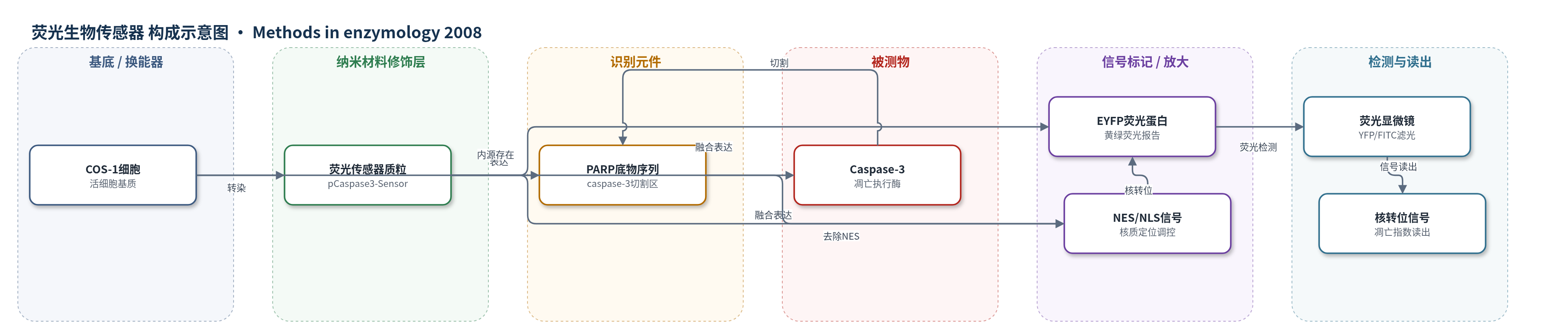
点击图片查看大图 · 依据论文自动绘制
传感器类型
荧光生物传感器
检测对象
Caspase-3 活性(caspase-3 activity)、细胞凋亡(apoptosis),样品基质:COS-1 活细胞(共转染 K-Ras 表达质粒)
检测原理
pCaspase3-Sensor 质粒在 COS-1 细胞中表达 EYFP-NES-PARP-NLS 融合蛋白。健康细胞中,NES 功能强于 NLS,使完整 EYFP 融合蛋白均匀分布于细胞质。当凋亡信号激活 caspase-3 时,caspase-3 切割 PARP 底物序列,移除 NES,生成 EYFP-NLS 片段;该片段借助 NLS 高效进入细胞核。因此,EYFP 荧光由细胞质向细胞核转移,核/质荧光比升高。荧光显微镜下,核阳性细胞比例(凋亡指数)随 caspase-3 活性和凋亡程度增加而升高。该方法无需外源底物或酶促放大,依靠基因编码荧光蛋白和核质转位实现单细胞读出。
检测灵敏度
原文未报告 LOD、线性范围、灵敏度斜率或相关系数。
效应效果
该 YFP-caspase 传感器可在单细胞水平区分细胞质与细胞核荧光,避免 Annexin-V、TUNEL 或 PARP 裂解等群体检测方法的平均化偏差。与目标基因共转染后,仅对表达 EYFP 的细胞评分,提高对转染细胞特异性的分析能力。COS-1 细胞转染效率通常超过 90%,且铺展良好,便于核转位判读;实验通常累计评分 100 个荧光模式清晰的细胞,计算凋亡指数。UV 辐射作为阳性对照可诱导核转位;K-Ras12V181E 诱导凋亡,K-Ras12V181A 不诱导,Bcl-2 过表达可阻断,显示对 caspase-3 依赖凋亡路径的响应。原文未报告 RSD、回收率或标准曲线。
传感器的构成
- 细胞基质:COS-1 细胞,提供活细胞表达与 caspase-3 激活环境
- 表达载体:pCaspase3-Sensor 质粒,编码 EYFP-NES-PARP-NLS 融合传感器
- 识别元件:PARP 底物序列(36 nt 编码区),被激活 caspase-3 特异性切割
- 信号标记物:EYFP(增强型黄绿色荧光蛋白),提供可检测荧光信号
- 定位调控元件:NES(核输出信号)与 NLS(核定位信号),控制 EYFP 在细胞质/细胞核分布
- 读出系统:荧光显微镜(YFP/FITC 滤光片),检测 EYFP 核转位并计算凋亡指数
中文摘要
K-Ras 通过 C 端法尼基异戊二烯修饰和邻近多碱性区与质膜结合,其膜定位可受多碱性区净电荷调节。蛋白激酶 C(PKC)磷酸化多碱性区 Ser181 可中和正电荷,从而调节法尼基-静电开关,控制 K-Ras 与质膜的结合;该开关激活还可促进凋亡。本章介绍多种方法:利用 32P 代谢标记、免疫沉淀、SDS-PAGE 和磷成像直接分析 K-Ras 磷酸化位点;通过 GFP 标记 K-Ras 的活细胞成像间接评估法尼基-静电开关及亚细胞转位;利用 Raf-1 的 Ras 结合域将 PKCγ 催化域靶向激活 K-Ras,以人工激活该开关;以及使用 YFP 标记的 caspase-3 生物传感器在单细胞水平检测凋亡。
英文摘要
K-Ras is a member of a family of proteins that associate with the plasma membrane by virtue of a lipid modification that inserts into the membrane and a polybasic region that associates with the anionic head groups of inner leaflet phospholipids. In the case of K-Ras, the lipid is a C-terminal farnesyl isoprenoid adjacent to a polylysine sequence. The affinity of K-Ras for the plasma membrane can be modulated by diminishing the net charge of the polybasic region. Among the ways this can be accomplished is phosphorylation by protein kinase C (PKC) of serine 181 within the polybasic region. Phosphorylation at this site regulates a farnesyl-electrostatic switch that controls association of K-Ras with the plasma membrane. Surprisingly, engagement of the farnesyl-electrostatic switch promotes apoptosis. This chapter describes methods for directly analyzing the phosphorylation status of K-Ras using metabolic labeling with (32)P, for indirectly assessing the farnesyl-electrostatic switch by following GFP-tagged K-Ras in live cells, for artificially activating the farnesyl-electrostatic switch by directing the kinase domain of a PKC to activated K-Ras using a Ras-binding domain, and for assessing apoptosis of individual cells using a YFP-tagged caspase 3 biosensor.