组成图示
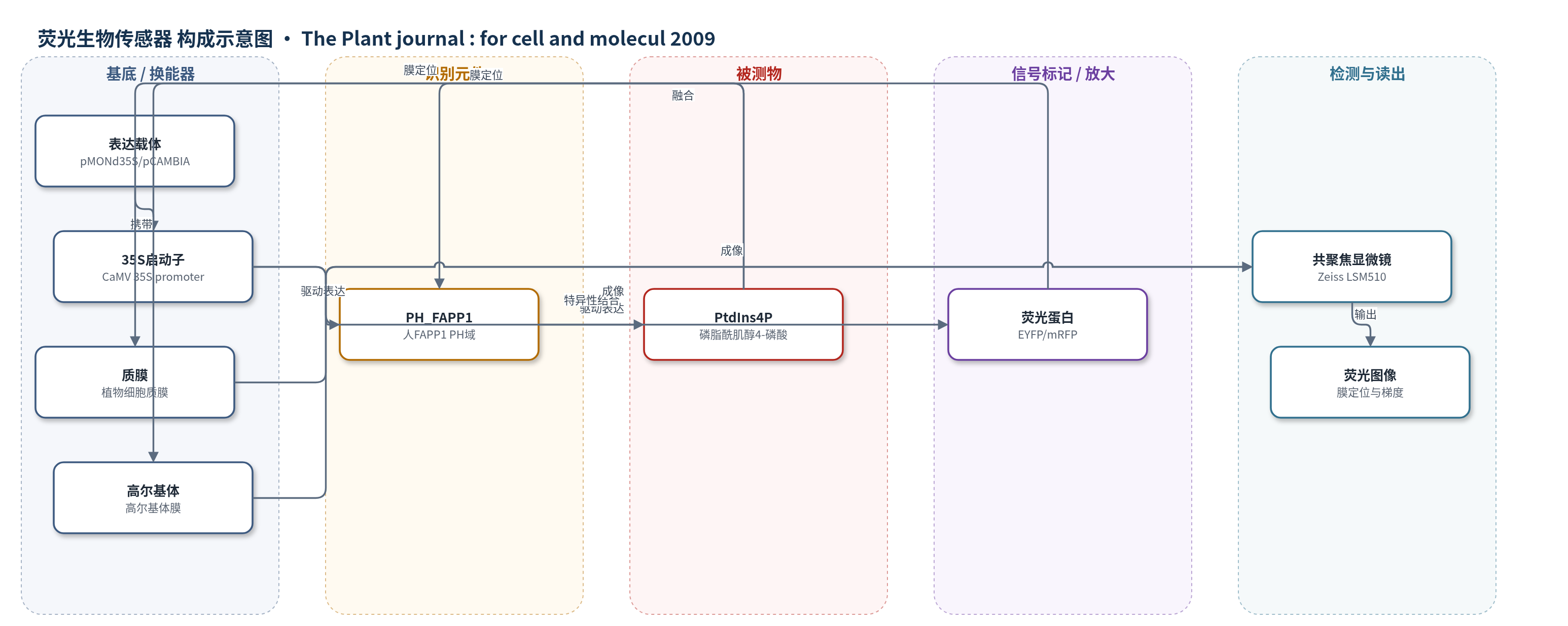
点击图片查看大图 · 依据论文自动绘制
传感器类型
荧光生物传感器
检测对象
磷脂酰肌醇4-磷酸(PtdIns4P);样品基质:豇豆原生质体、烟草 BY-2 细胞、Medicago truncatula 根、Arabidopsis thaliana 幼苗等活植物细胞/组织
检测原理
该传感器为荧光蛋白与脂质结合域的融合蛋白。PH_FAPP1特异性识别并结合膜脂PtdIns4P,使原本可扩散于胞质和核中的YFP/mRFP-PH_FAPP1被招募到富含PtdIns4P的膜界面,如质膜和高尔基体膜。PtdIns4P局部丰度越高,膜结合态传感器越多,相应膜上的荧光越强;未结合态呈胞质/核背景。共聚焦显微镜激发EYFP或mRFP并采集荧光图像,从而实时显示PtdIns4P的亚细胞定位、动态变化和浓度梯度。K7E/R18L突变破坏PH_FAPP1与PtdIns4P的结合,使膜定位消失,证明信号依赖识别事件。该法无酶促或核酸放大,直接以荧光蛋白作为换能信号。
检测灵敏度
原文未报告 LOD、线性范围、灵敏度斜率或相关系数。
效应效果
该传感器在豇豆原生质体、烟草BY-2细胞、Medicago truncatula根和Arabidopsis thaliana幼苗中表达,不影响细胞生长。K7E和R18L突变体丧失质膜定位;与STtmd-CFP强共定位,不与GFP-AtRABF2b共定位,且与YFP-2·FYVE不重叠。20 μM PAO处理20 min使mRFP-PH_FAPP1膜定位丢失,YFP-2·FYVE几乎不变;10 μM wortmannin处理20 min使YFP-2·FYVE点状结构丢失,mRFP-PH_FAPP1不受影响,30 μM亦无改变。50 μM BFA处理30 min形成大BFA区室,洗脱可逆。rhd4-1突变体PtdIns4P约增加50%,传感器主要定位内膜。生长根毛尖端出现PtdIns4P梯度,非生长根毛无,适用于活植物细胞PtdIns4P动态成像。
传感器的构成
- 表达载体:pMONd35S 或 pCAMBIA-35S 载体,含 CaMV 35S 启动子,驱动传感器在植物细胞中表达
- 荧光换能元件:EYFP 或 mRFP 荧光蛋白,提供共聚焦显微镜可检测的荧光信号
- 识别元件:人 FAPP1 蛋白的 PH 结构域(PH_FAPP1),特异性结合 PtdIns4P
- 融合传感器:YFP-PH_FAPP1 或 mRFP-PH_FAPP1,将识别域与荧光蛋白融合,实现 PtdIns4P 膜定位可视化
- 表达基质:豇豆原生质体、烟草 BY-2 细胞、Medicago truncatula 根、Arabidopsis thaliana 幼苗,作为活细胞成像样品
- 特异性对照:PH_FAPP1-K7E 或 PH_FAPP1-R18L 突变体,破坏 PtdIns4P 结合,用于验证定位特异性
中文摘要
多磷酸肌醇是含量低于总磷脂1%的少数磷脂,却参与多种信号转导和膜运输事件。磷脂酰肌醇4-磷酸(PtdIns4P)是最丰富的多磷酸肌醇,周转迅速,但其在细胞或植物中的具体分布仍不清楚。本文报道了一种用于监测活植物细胞中PtdIns4P动态的脂质生物传感器。该传感器由荧光蛋白与特异性结合PtdIns4P的脂质结合域融合而成,即人FAPP1蛋白的PH结构域(PH_FAPP1)。YFP-PH_FAPP1在豇豆原生质体瞬时表达,并在烟草BY-2细胞、Medicago truncatula根和Arabidopsis thaliana幼苗中稳定表达,均无明显不利影响。观察到两种荧光模式:运动点状结构和质膜。共表达标记显示,该传感器与高尔基体标记STtmd-CFP强共定位,不与内吞/前液泡标记GFP-AtRABF2b共定位;与PtdIns3P传感器YFP-2·FYVE定位完全不同。细胞分裂时,YFP-PH_FAPP1强烈标记细胞板,而PtdIns3P缺失于新形成的细胞膜。在Medicago和Arabidopsis生长根毛中,质膜出现明显的PtdIns4P梯度,尖端浓度最高,提示PtdIns4P参与尖端生长。
英文摘要
Polyphosphoinositides represent a minor group of phospholipids, accounting for less than 1% of the total. Despite their low abundance, these molecules have been implicated in various signalling and membrane trafficking events. Phosphatidylinositol 4-phosphate (PtdIns4P) is the most abundant polyphosphoinositide. (32)Pi-labelling studies have shown that the turnover of PtdIns4P is rapid, but little is known about where in the cell or plant this occurs. Here, we describe the use of a lipid biosensor that monitors PtdIns4P dynamics in living plant cells. The biosensor consists of a fusion between a fluorescent protein and a lipid-binding domain that specifically binds PtdIns4P, i.e. the pleckstrin homology domain of the human protein phosphatidylinositol-4-phosphate adaptor protein-1 (FAPP1). YFP-PH(FAPP1) was expressed in four plant systems: transiently in cowpea protoplasts, and stably in tobacco BY-2 cells, Medicago truncatula roots and Arabidopsis thaliana seedlings. All systems allowed YFP-PH(FAPP1) expression without detrimental effects. Two distinct fluorescence patterns were observed: labelling of motile punctate structures and the plasma membrane. Co-expression studies with organelle markers revealed strong co-labelling with the Golgi marker STtmd-CFP, but not with the endocytic/pre-vacuolar marker GFP-AtRABF2b. Co-expression with the Ptdins3P biosensor YFP-2 x FYVE revealed totally different localization patterns. During cell division, YFP-PH(FAPP1) showed strong labelling of the cell plate, but PtdIns3P was completely absent from the newly formed cell membrane. In root hairs of M. truncatula and A. thaliana, a clear PtdIns4P gradient was apparent in the plasma membrane, with the highest concentration in the tip. This only occurred in growing root hairs, indicating a role for PtdIns4P in tip growth.