组成图示
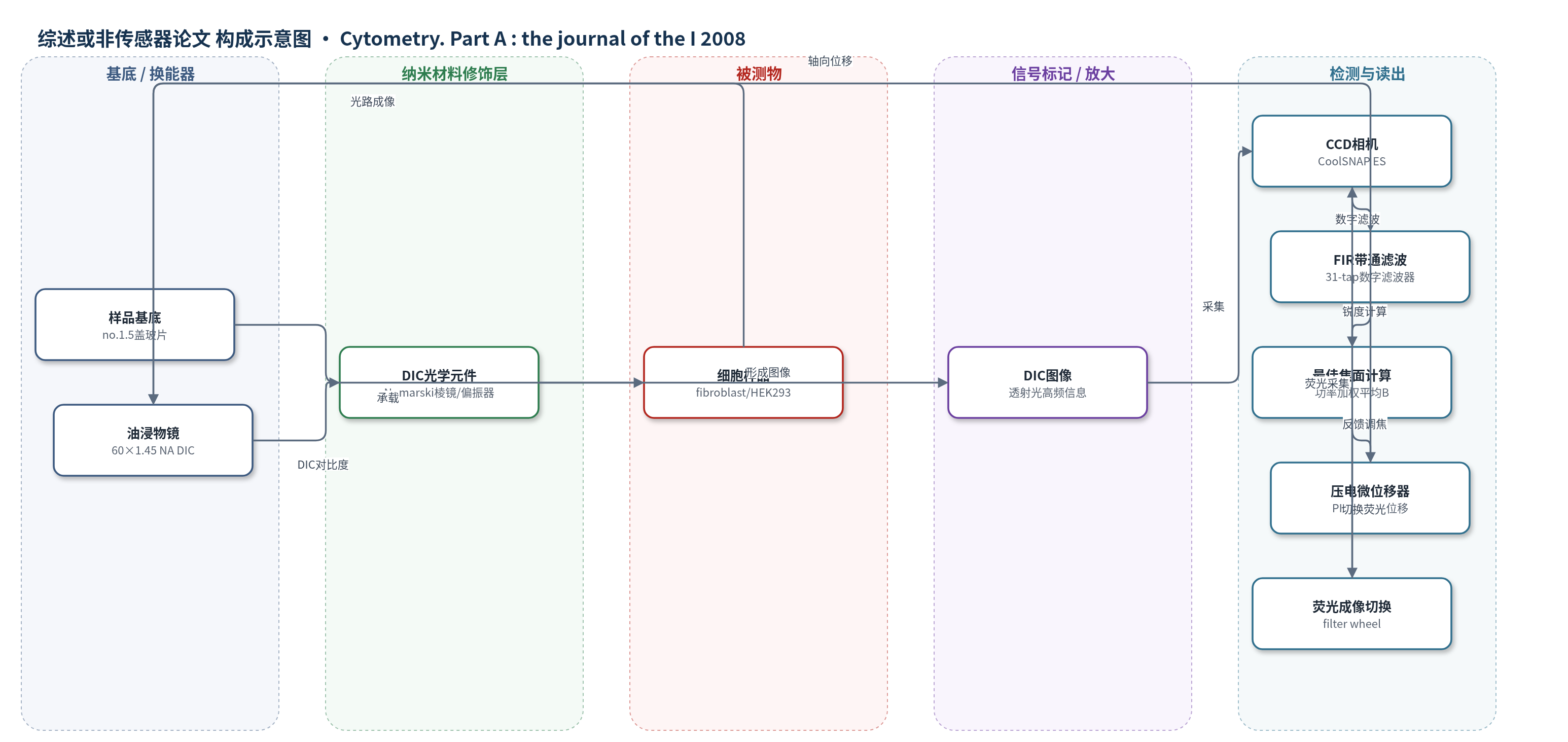
点击图片查看大图 · 依据论文自动绘制
传感器类型
综述或非传感器论文
检测对象
细胞样品(mouse embryonic fibroblasts、HEK 293 cells),样品基质:no.1.5盖玻片/玻璃载玻片上的固定或活细胞培养物;非生物传感分析物。
检测原理
本文不是基于识别元件与信号标记物的浓度响应传感,而是基于DIC透射光成像的自动对焦。样品离焦时,图像高频空间频率功率下降;系统用31-tap FIR带通滤波器提取归一化频率0.12–0.2的高频带,计算每个z平面的锐度F(z),并以功率加权平均得到最佳焦面B。PIFOC压电微位移器按B移动物镜,重复采集与计算,直至达到最佳焦面。低粘度油浸介质(135 cSt)和50 ms延迟用于消除油层黏滞导致的位移滞后。因此信号随离焦程度变化,而不随生物分子浓度变化。
检测灵敏度
自动对焦精度: 8.6 nm (SD, mean over 225 FOVs);combined SD: 10.3 nm;单视野SD范围: 2.2–34.4 nm;相关系数: r^2 = 0.97(细胞厚度与铺板密度);r^2 = 0.28(自动对焦SD与厚度)。
效应效果
在225个视野、11,250次自动对焦试验中,最佳焦面标准差平均为8.6 nm,合并标准差为10.3 nm,明显优于此前20×0.75 NA相衬系统的56 nm。改变铺板密度使细胞厚度从9.47 μm增至33.20 μm时,自动对焦标准差无显著趋势(r^2=0.28),说明精度与样品厚度基本无关。使用135 cSt低粘度油并设50 ms延迟后,2 μm焦程、500 nm步长可在1.0 s内完成自动对焦。96孔板玻璃底初步实验在8×4孔区域有效。该方法避免相衬弯月面畸变,并减少额外荧光曝光,适用于高分辨率活细胞延时成像。
传感器的构成
- 样品基底:no.1.5盖玻片/玻璃载玻片,承载固定或活细胞样品。
- 样品/被测对象:mouse embryonic fibroblasts、HEK 293 cells,提供DIC成像结构。
- 成像物镜:60×1.45 NA PlanApo DIC TIRF油浸物镜,收集透射光并生成高空间频率信息。
- DIC光学元件:Nomarski棱镜与线性偏振器,产生微分干涉差对比度。
- 轴向位移换能器:PIFOC压电微位移器(Physik Instrumente 7-721.10),移动物镜实现z方向对焦。
- 图像采集器:Photometrics CoolSNAP ES CCD相机,采集DIC图像。
- 数字信号处理:31-tap FIR带通滤波器与Visual C++程序,提取高频锐度并计算最佳焦面。
中文摘要
随着细胞荧光生物传感器的发展,单活细胞内蛋白动态研究日益重要。自动对焦可补偿温度漂移、多视野基底不平整以及长时间延时成像中的细胞生长,对数小时至数天的高分辨率研究尤为关键。为获得最高分辨率与灵敏度,常需使用高数值孔径油浸物镜,同时限制荧光曝光以降低光毒性。由于相衬成像在微孔板中受弯月面畸变影响,作者研究了移除荧光光路分析器后,采用60×1.45 NA油浸物镜的微分干涉差(DIC)自动对焦。基于实验DIC调制传递函数,设计了一种新的带通数字滤波器用于测量图像锐度。在225个视野的重复测试中,自动对焦精度为8.6 nm(标准差)。通过改变细胞铺板密度控制样品厚度(9.47–33.20 μm),结果显示自动对焦精度与样品厚度无关。该结果表明,所选空间频率可实现高NA DIC自动显微术的高精度自动对焦,有望消除微孔板相衬成像中的弯月面畸变,并避免额外荧光曝光带来的毒性。
英文摘要
Continued advances in cellular fluorescent biosensors enable studying intracellular protein dynamics in individual, living cells. Autofocus is valuable in such studies to compensate for temperature drift, uneven substrate over multiple fields of view, and cell growth during long-term high-resolution time-lapse studies of hours to days. Observing cellular dynamics with the highest possible resolution and sensitivity motivates the use of high numerical aperture (NA) oil-immersion objectives, and control of fluorescence exposure to minimize phototoxicity. To limit phototoxicity, to maximize light throughput of the objective for biosensor studies, and because phase contrast is distorted by the meniscus in microtiter plates, we studied autofocus in differential interference contrast (DIC) microscopy with a 60x 1.45 NA oil objective after removing the analyzer from the fluorescent light path. Based on a study of the experimental DIC modulation transfer function, we designed a new bandpass digital filter for measuring image sharpness. Repeated tests of DIC autofocus with this digital filter on 225 fields-of-view resulted in a precision of 8.6 nm (standard deviation). Autofocus trials on specimens with thicknesses from 9.47 to 33.20 mum, controlled by cell plating density, showed that autofocus precision was independent of specimen thickness. The results demonstrated that the selected spatial frequencies enabled very high-precision autofocus for high NA DIC automated microscopy, thereby potentially removing the problems of meniscus distortion in phase contrast imaging of microtiter plates and rendering the toxicity of additional fluorescence exposure unnecessary.