组成图示
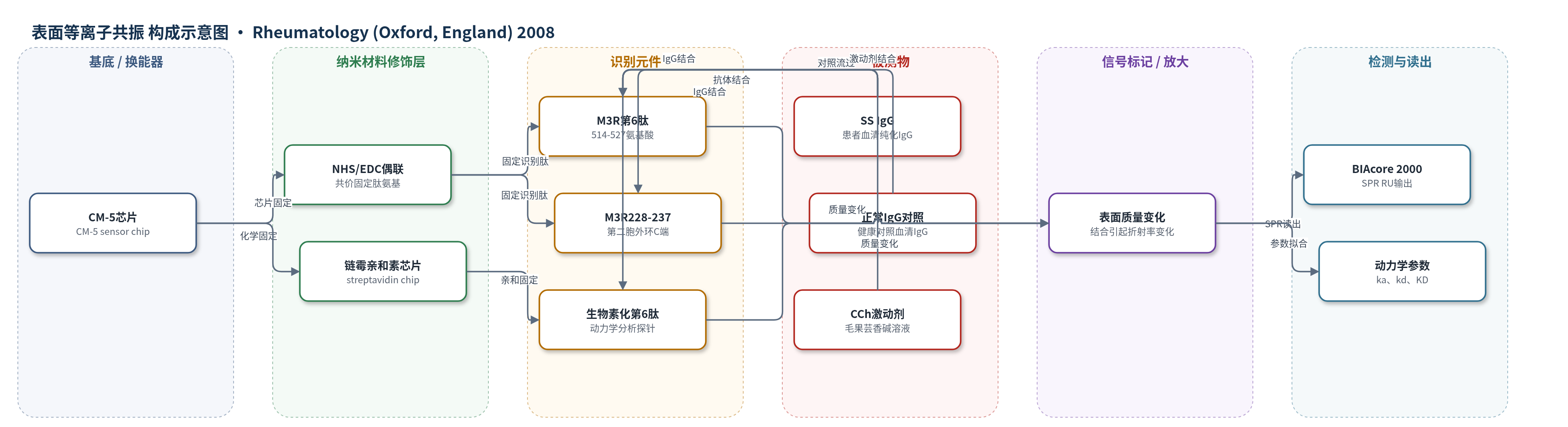
点击图片查看大图 · 依据论文自动绘制
传感器类型
表面等离子共振(SPR)生物传感器
检测对象
干燥综合征(Sjögren’s syndrome, SS)患者血清纯化免疫球蛋白 G(IgG,抗 M3R 自身抗体)、正常对照血清 IgG、毛果芸香碱(carbachol, CCh);样品基质为血清纯化 IgG 稀释液/SPR 流动相。
检测原理
该检测基于表面等离子共振(SPR)直接监测分子结合。首先用 NHS/EDC 胺偶联将合成 M3R 肽(如第6肽 514–527、M3R228–237)共价固定于 CM-5 芯片表面;也可用链霉亲和素芯片固定生物素化第6肽。将不同浓度 SS IgG、正常 IgG 或 CCh 在 running buffer 中流过芯片,若分析物与表面肽特异性结合,界面质量/折射率增加,SPR 共振角或波长偏移,BIAcore 2000 记录为共振单位(RU)上升;洗脱后结合物解离,RU 回落。结合量随 IgG 或 CCh 浓度增加而增加,形成浓度依赖传感器图。按 1:1 Langmuir 模型拟合可得到结合速率常数 ka、解离速率常数 kd 和表观解离常数 KD。该方法无酶或荧光放大,信号直接来自抗原-抗体/配体-受体结合。
检测灵敏度
原文未报告 LOD、线性范围、灵敏度斜率或相关系数。
效应效果
HSG 细胞中,0.5 mg/ml SS IgG 使 CCh 诱导胞内钙瞬变(CICT)抑制 94.8±0.65%(0.012±0.001,n=9,P<0.05);ATP 诱导钙信号仍为 0.156±0.008,与正常 IgG 组 0.154±0.004 无显著差异,提示特异性抑制 M3R。第6肽与 SS IgG 共孵育后 CICT 恢复至 0.233±0.011(n=11),约 97.07±5%(n=12)。BIAcore 显示第4肽和第6肽与 SS IgG 浓度依赖结合,1200 nM 时分别为 35.0±0.10 和 91.66±0.77 RU;正常 IgG 不与第6肽结合,但可与第4肽非特异结合。第6肽与 SS IgG 的 KD 为 3.87±2.2 μM(n=3)。7 例 SS 患者中仅部分原发性 SS IgG 结合第6肽,SS43 结合 M3R228–237。作者认为第三胞外环是功能表位和潜在治疗靶点。
传感器的构成
- 基底/换能器:CM-5 sensor chip(SPR 芯片)提供肽固定表面与光学传感界面
- 化学修饰层:NHS/EDC 胺偶联试剂将肽氨基共价固定到芯片表面
- 识别元件:合成 M3R 肽(第6肽 514–527、M3R228–237、第4肽等)作为表位探针
- 替代固定层:streptavidin sensor chip 与生物素化第6肽,用于动力学分析
- 流动相:running buffer(150 mM NaCl、10 mM HEPES、3 mM EDTA、0.005% surfactant P20,pH 7.4)维持结合环境
- 还原剂:1 mM DTT 保持第6肽二硫键还原状态
- 分析物:SS IgG、正常 IgG 或 CCh 作为待测结合分子
- 信号读出:BIAcore 2000 检测 SPR 共振单位(RU)随结合/解离变化
中文摘要
近年来,干燥综合征(Sjögren’s syndrome, SS)患者血清中可检出针对毒蕈碱型 M3 受体(M3R)的自身抗体,但与 SS 免疫球蛋白 G(IgG)相互作用的 M3R 精确表位仍不清楚。本研究旨在鉴定与 SS IgG 结合并影响 M3R 功能的功能性表位。作者从 7 例 SS 患者(6 例原发性、1 例继发性)和 2 例正常对照血清中纯化 IgG,检测 SS IgG 是否抑制 M3R 功能,并利用覆盖 M3R 全部胞外区的 6 条合成肽,通过微分光光度法和基于表面等离子共振(SPR)的光学生物传感器系统(BIAcore)鉴定表位。结果显示,0.5 mg/ml SS IgG 可抑制人下颌下腺(HSG)细胞中毛果芸香碱(CCh)诱导的胞内钙瞬变(CICT);与第 6 条肽(514–527 氨基酸区,对应 M3R 第三胞外环)共孵育后 CICT 恢复,BIAcore 进一步证实第 6 肽与 3 例原发性 SS 患者 IgG 呈浓度依赖性结合。对应 M3R 第二胞外环 C 端 228–237 氨基酸的肽也结合 SS IgG,而正常 IgG 不与第 6 肽结合。结果表明,M3R 第三胞外环是 SS IgG 结合的功能性表位,并部分抑制 M3R 功能。
英文摘要
OBJECTIVES: Recently, autoantibodies directed against muscarinic type 3 receptor (M3R) have been reported in patients with primary SS. However, the precise epitope(s) of the M3R that interacts with SS autoantibodies remains unclear. The aim of this study was to identify the functional epitope of M3R which interacts with SS immunoglobulin G (IgG).
METHODS: Purified IgGs were obtained from the sera of seven SS patients (six primary and one secondary SS) and two normal persons. We examined whether SS IgG inhibits M3R function and identified the epitope using six synthetic peptides covering all the extracellular domains of M3R by microspectrofluorimetry and surface plasmon resonance-based optical biosensor system (BIAcore system).
RESULTS: A volume of 0.5 mg/ml SS IgG inhibited carbachol (CCh)-induced [Ca(2+)](i) transient (CICT) in human submandibular gland (HSG) cells. However, co-incubation of SS IgG with the 6th peptide (514-527 amino acid region) corresponding to the third extracellular loop of M3R, recovered CICT. The result was further confirmed by BIAcore analysis. We found that the 6th peptide interacts with IgGs from three primary SS patients in a concentration-dependent manner. The synthetic peptide which consists of amino acids 228-237 corresponding to the COOH-terminus of the second extracellular loop of M3R also bound to SS IgG. However, normal IgGs did not interact with the 6th peptide.
CONCLUSIONS: The results suggest that the third extracellular loop of M3R represents a functional epitope bound by SS IgG, and thereby partly inhibits M3R function.