组成图示
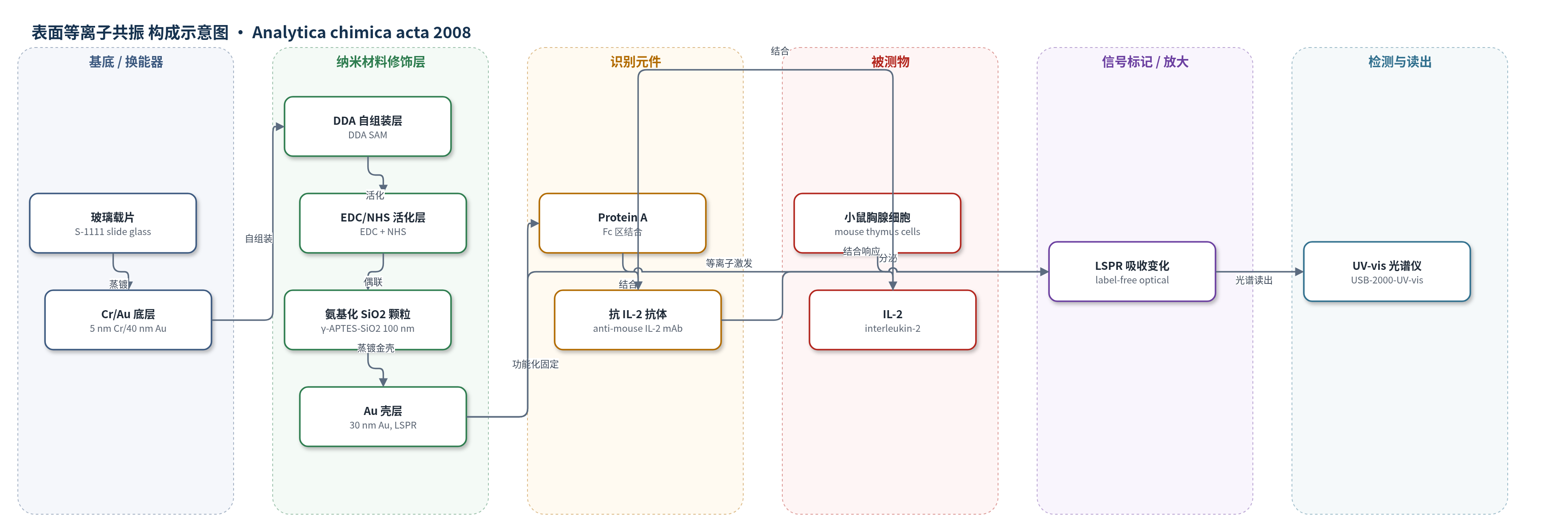
点击图片查看大图 · 依据论文自动绘制
传感器类型
表面等离子共振(SPR)生物传感器
检测对象
白细胞介素-2(IL-2,interleukin-2);样品基质:小鼠胸腺分离细胞在 20 mM PBS 中经 Con-A 刺激后的细胞代谢上清/校准 IL-2 溶液
检测原理
该传感器以 Au 壳层包覆的 SiO2 纳米颗粒层作为 LSPR 换能界面。抗小鼠 IL-2 抗体经 Protein A 固定于传感器表面。当小鼠胸腺细胞经 Con-A 刺激后分泌 IL-2 时,IL-2 与表面抗体特异性结合,使金壳层附近生物分子层厚度及局部折射率增加。Au 纳米壳的局域表面等离子共振吸收峰对界面折射率变化敏感,因此 LSPR 吸收强度随 IL-2 浓度增加而增大。系统通过 UV–vis 光谱监测吸收峰变化,无需酶或荧光标记,即可实现细胞代谢物的无标记定量与时间序列分析。
检测灵敏度
LOD: 10 pg mL−1;校准范围: 0–100 μg mL−1(半对数响应)
效应效果
该传感器对 IL-2 具有免疫特异性:100 μg/mL BSA 阴性对照仅引起约 0.01 a.u. 非特异吸附背景;未加 Con-A 刺激或使用抗 DNP-BSA 抗体时未观察到显著吸收变化(约 0.05–0.01 a.u.)。Con-A 刺激小鼠胸腺细胞后,测得 IL-2 代谢量约为 75–150 pg/mL,并可实现 1–48 h 时间序列监测。与 ELISPOT 相比,LSPR 方法可提供定量浓度信息;与 ELISA 相比,其校准行为与代谢趋势基本一致,且无需标记、装置更简单、成本低于传统 SPR,适合高通量细胞活性分析。
传感器的构成
- 基底/换能器电极:玻璃载片(S-1111)上热蒸镀 5 nm Cr 和 40 nm Au,提供附着基底并支撑后续纳米颗粒层。
- 自组装单分子层:4,4'-二硫代丁酸(DDA)在 Au 表面形成羧基端 SAM,用于 EDC/NHS 活化及颗粒/抗体固定。
- 活化/交联层:EDC 活化 DDA 羧基,NHS 参与形成反应性酯,用于连接氨基化 SiO2 颗粒和 Protein A/抗体。
- 纳米颗粒核:γ-APTES 修饰的 100 nm 二氧化硅(SiO2)纳米颗粒,表面氨基与活化 DDA 偶联形成颗粒层。
- 金壳层:热蒸镀 30 nm Au 覆盖 SiO2 颗粒,形成核–壳结构并激发局域表面等离子共振(LSPR)。
- 识别元件:Protein A 结合 IgG Fc 区,抗小鼠 IL-2 单克隆抗体(anti-IL-2 mAb)特异性识别 IL-2。
- 信号标记物:无标记检测,不引入酶、荧光或化学发光标记,信号来自抗原–抗体结合引起的 LSPR 光学变化。
- 光学读出:UV–vis 光谱仪(USB-2000-UV–vis)、钨卤素光源和光纤探头监测 LSPR 吸收峰变化。
中文摘要
为理解生命活动,细胞作为最小生命单元的分析对生物学与生物技术具有重要意义。本研究提出一种基于局域表面等离子共振(LSPR)生物传感器的无标记细胞检测方法,该传感器采用核–壳结构纳米颗粒层作为激发基底。为验证其性能,作者利用小鼠胸腺分离细胞检测细胞代谢物。在固定抗体的 LSPR 传感器表面,通过抗原–抗体特异性相互作用引起的折射率变化来检测细胞代谢物,并监测抗体与细胞代谢物之间特异性反应的光学特性。结果显示,该抗体固定 LSPR 生物传感器的检出限为 10 pg/mL,并实现了小鼠胸腺分离细胞代谢过程的时间序列分析。结果表明,LSPR 生物传感器为低成本、简化装置和小样品量条件下检测生物分子相互作用提供了有前景的平台。
英文摘要
For an understanding of the life activities, the analysis of cells, which are the smallest units of life form, has a significant impact on biology and biotechnology. In this study, we propose a novel label-free cell-based assay that is based on localized surface plasmon resonance (LSPR) biosensor, which is excited using core-shell structured nanoparticle layer substrate. To demonstrate the promising properties of our LSPR-based label-free cell-based assay, we performed the detection of the cell metabolites using the isolated cells from mouse thymus. For detection of the cellular metabolites, the refractive index change by the specific interaction between the antigen and antibody was detected on the antibody immobilized LSPR-based biosensor. Using our LSPR-based biosensor, the optical characteristics were monitored for the detection of specific reactions between antibody and cell metabolites. As a result, the detection limit of this antibody immobilized LSPR-based biosensor was 10 pg mL(-1). Furthermore, the time-course analysis of cell metabolisms using the isolated cells from mouse thymus was also achieved. From these results, the LSPR-based biosensor provides a promising platform with attractive advantages for the detection of biomolecular interactions at low-cost in a simplified experimental set-up with a low sample volume.