组成图示
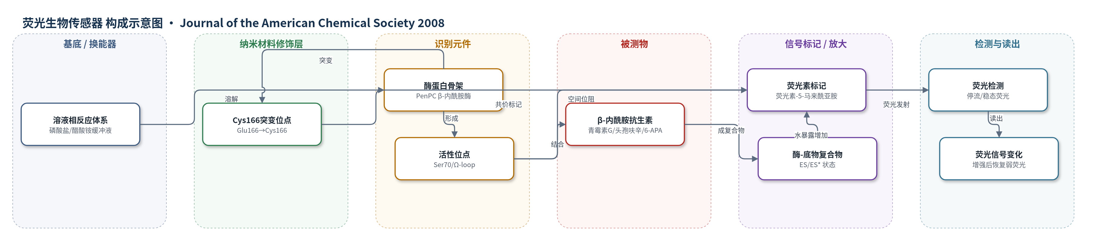
点击图片查看大图 · 依据论文自动绘制
传感器类型
荧光生物传感器
检测对象
青霉素 G(penicillin G)、头孢呋辛(cefuroxime)、6-氨基青霉烷酸(6-APA);样品基质为磷酸盐缓冲液或醋酸铵缓冲液溶液
检测原理
E166Cf 由 PenPC β-内酰胺酶的 Glu166 突变为 Cys166 并共价连接环境敏感荧光素-5-马来酰亚胺构成。游离酶状态下,荧光素埋藏于活性位点附近,溶剂可及面积较低,荧光较弱。β-内酰胺抗生素进入活性位点后,与 Ser70 等残基结合形成非共价 ES 复合物,并进一步酰化形成共价 ES* 复合物。抗生素的 β-内酰胺环和噻唑烷环与荧光素占据共同空间,产生空间位阻,迫使荧光素从活性位点移向外部水相,溶剂可及面积由约191 Ų增至约350 Ų,水暴露增加使荧光增强。若底物被缓慢水解,酶再生为游离态,荧光素返回活性位点,荧光恢复弱信号。因此荧光强度随酶-底物复合物比例增加而增强,可反映 β-内酰胺抗生素的结合与存在。
检测灵敏度
原文未报告 LOD、线性范围、灵敏度斜率或相关系数。
效应效果
该传感器可识别 β-内酰胺抗生素,青霉素 G、头孢呋辛和 6-APA 均引起荧光增强;6-APA 仅含双环结构也能响应,说明 β-内酰胺环/噻唑烷环是触发荧光素位移的关键。E166Cf 与头孢呋辛 Kd=0.91±0.09 mM、kcat=2.0±0.2×10^-4 s^-1;与 6-APA Kd=0.7±0.1 mM、kcat=8.0±1.0×10^-4 s^-1;与青霉素 G Km=213±11 µM、kcat=5.28±0.09 s^-1。热变性 Tm 为野生型 56±1 °C、E166C 50±1 °C、E166Cf 49±1 °C,突变降低稳定性但增加 Ω-loop 柔性,可补偿荧光素位阻。作者认为该模型可用于临床诊断、环境监测和食品抗生素滥用检测。
传感器的构成
- 识别/换能蛋白:PenPC β-内酰胺酶 E166C 突变体(E166Cf),Glu166→Cys166,结合并缓慢水解 β-内酰胺抗生素,提供识别与微环境变化
- 信号标记物:荧光素-5-马来酰亚胺(fluorescein-5-maleimide),共价连接 Cys166,作为环境敏感荧光探针,水暴露增加时荧光增强
- 识别位点:酶活性中心(含 Ser70 等残基),与 β-内酰胺环/噻唑烷环结合,形成 ES 和 ES* 复合物
- 反应介质:磷酸盐缓冲液(50 mM potassium phosphate, pH 7.0)或醋酸铵缓冲液(20 mM ammonium acetate, pH 7.0),维持酶构象与反应
中文摘要
荧光素标记的 PenPC β-内酰胺酶 E166C 突变体(E166Cf)是从非变构蛋白构建“开关型”荧光生物传感器的成功模型。本文重点研究 E166Cf 在 β-内酰胺抗生素结合与水解过程中荧光变化的传感机制。利用电喷雾质谱和停流荧光研究 E166Cf 与头孢呋辛、青霉素 G 及 6-氨基青霉烷酸(6-APA)的相互作用,发现酶-底物复合物的形成可增强 E166Cf 的荧光,而随后游离酶的再生使其恢复弱荧光。E166Cf 与青霉素 G 的分子建模显示,荧光素标记很可能与抗生素的 β-内酰胺环和噻唑烷环在活性位点占据共同空间;这种空间位阻使荧光素在底物结合时从活性位点移向外部水相环境,从而暴露于更高水环境。稳态荧光测量表明,仅含 β-内酰胺环和噻唑烷环的 6-APA 也能增强 E166Cf 荧光。对野生型酶、E166C 和 E166Cf 的热变性实验表明,E166C 突变可能增加 Ω-loop 的柔性,这种结构改变可补偿荧光素标记对底物结合可能产生的空间位阻效应。
英文摘要
The fluorescein-labeled E166C mutant of the PenPC beta-lactamase (E166Cf) represents a successful model in the construction of "switch-on" fluorescent biosensors from nonallosteric proteins (Chan P.-H. et al.; J. Am Chem. Soc., 2004, 126, 4074). This paper focuses on the study of the biosensing mechanism by which the E166Cf biosensor changes its fluorescence upon beta-lactam binding and hydrolysis. Mass spectrometric and stopped-flow fluorescence studies of E166Cf with cefuroxime, penicillin G, and 6-aminopenicillanic acid reveal that the formation of enzyme-substrate complex enhances the fluorescence of E166Cf, and the subsequent regeneration of the free enzyme restores the weak fluorescence of E166Cf. Molecular modeling studies of E166Cf with penicillin G show that the fluorescein label is likely to share a common space with the beta-lactam and thiazolidine rings of the antibiotic in the active site. This spatial clash appears to cause the fluorescein label to move from the active site to the external aqueous environment upon substrate binding and hence experience higher water exposure. Steady-state fluorescence measurements indicate that the fluorescence of E166Cf can be enhanced by 6-aminopenicillanic acid, which consists of the beta-lactam and thiazolidine rings only. Thermal denaturation experiments of the wild-type enzyme, E166C, and E166Cf reveal that the E166C mutation is likely to increase the flexibility of the Omega-loop. This "modified" structural property might compensate for the possible steric effect of the fluorescein label on substrate binding.