组成图示
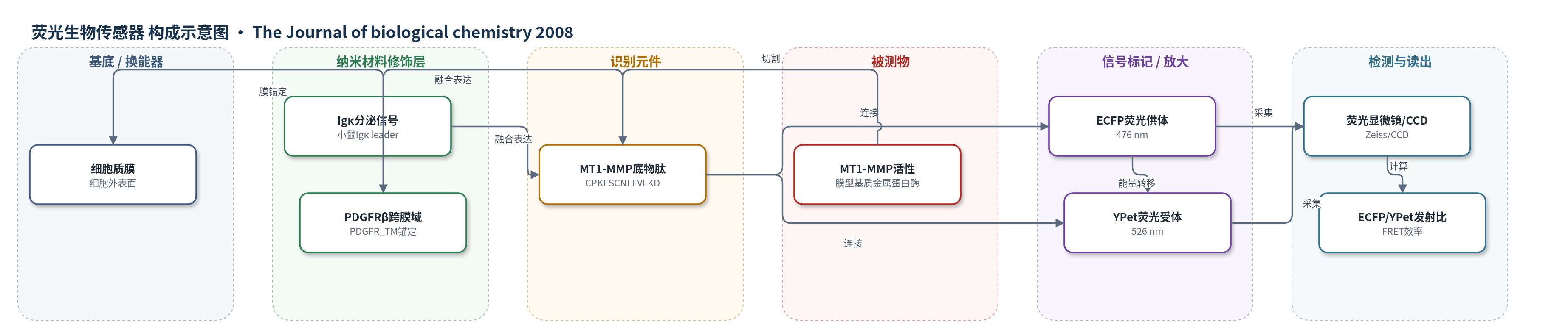
点击图片查看大图 · 依据论文自动绘制
传感器类型
荧光生物传感器
检测对象
膜型1基质金属蛋白酶活性(MT1-MMP activity),样品基质为活细胞(HeLa、MDA-MB-231、HT-1080、成纤维细胞等细胞培养体系)
检测原理
该传感器将MT1-MMP敏感底物肽CPKESCNLFVLKD连接在ECFP与YPet之间,并通过PDGFRβ跨膜域锚定于细胞外表面。当膜型MT1-MMP活性升高时,其催化域在细胞表面切割底物肽,使ECFP与YPet空间分离,二者距离超过FRET有效范围,FRET效率下降。激发ECFP后,原本转移给YPet的非辐射能量减少,导致476 nm ECFP发射增强、526 nm YPet发射减弱,ECFP/YPet发射比升高。荧光显微镜逐像素采集双通道荧光并计算比值,即可将MT1-MMP活性映射为FRET信号;活性越高,切割比例越大,比值越高。EGF通过EGFR信号募集并激活MT1-MMP,使局部切割增强,从而在迁移前缘形成方向性FRET响应。
检测灵敏度
原文未报告LOD、线性范围、灵敏度斜率或相关系数。
效应效果
体外实验中,传感器对MT1-MMP催化域(2 μg/ml)响应强,对MT3-MMP也有响应,而对MT2-MMP、MMP-2和MMP-9(6 μg/ml)基本无响应。活细胞中,EGF(50 ng/ml)刺激60 min或3 h可诱导显著ECFP/YPet比升高;MT1-MMP缺陷细胞无响应,野生型MT1-MMP可恢复,MMP-2、MMP-9及无活性突变体不能。TIMP-2(2.5 μg/ml)和GM6001抑制响应,TIMP-1不抑制,显示对膜型MMP的选择性。细胞骨架破坏剂Cyto D或nocodazole削弱方向性响应。作者认为该传感器可用于活细胞亚细胞分辨率成像和MT1-MMP抑制剂高通量筛选。
传感器的构成
- 荧光供体:ECFP(enhanced cyan fluorescent protein),作为FRET供体,切割后476 nm荧光增强
- 荧光受体:YPet,作为FRET受体,与ECFP形成高动态范围FRET对
- 识别底物:MT1-MMP敏感肽CPKESCNLFVLKD,来源于proMMP-2切割位点,被MT1-MMP切割
- 分泌信号:小鼠Igκ链leader sequence,引导传感器进入分泌途径并暴露于细胞外
- 膜锚定:PDGFRβ跨膜域(PDGFR_TM),将传感器靶向并锚定于质膜
- 表达载体:pDisplay质粒,用于哺乳动物细胞表达膜靶向FRET传感器
- 信号读出:倒置荧光显微镜与CCD相机,采集ECFP/YPet发射比(476/526 nm)
中文摘要
膜型1基质金属蛋白酶(MT1-MMP)通过蛋白水解重塑细胞外基质,在癌细胞生物学中发挥关键作用。本研究利用荧光共振能量转移(FRET)成像技术开发了一种新型生物传感器,其传感元件锚定于细胞膜细胞外表面,可在活细胞中以亚细胞分辨率动态可视化 MT1-MMP 活性。表皮生长因子(EGF)在表达 MT1-MMP 的癌细胞中诱导显著 FRET 变化,但在 MT1-MMP 缺陷细胞中不诱导;该变化可被野生型 MT1-MMP 恢复,但不能被 MMP-2、MMP-9 或无活性 MT1-MMP 突变体恢复。删除传感器跨膜域或用细胞不可渗透抑制剂 TIMP-2 处理可消除 EGF 诱导的 FRET 响应,表明 MT1-MMP 在细胞表面产生信号。EGF 刺激下,活性 MT1-MMP 被定向募集至沿微图案纤连蛋白条纹迁移细胞的前缘,并与 EGFR 局部积累同步,该过程依赖完整细胞骨架网络。该传感器为研究关键蛋白水解酶时空调控机制提供了有力工具。
英文摘要
Membrane type 1 matrix metalloproteinase (MT1-MMP) plays a critical role in cancer cell biology by proteolytically remodeling the extracellular matrix. Utilizing fluorescence resonance energy transfer (FRET) imaging, we have developed a novel biosensor, with its sensing element anchoring at the extracellular surface of cell membrane, to visualize MT1-MMP activity dynamically in live cells with subcellular resolution. Epidermal growth factor (EGF) induced significant FRET changes in cancer cells expressing MT1-MMP, but not in MT1-MMP-deficient cells. EGF-induced FRET changes in MT1-MMP-deficient cells could be restored after reconstituting with wild-type MT1-MMP, but not MMP-2, MMP-9, or inactive MT1-MMP mutants. Deletion of the transmembrane domain in the biosensor or treatment with tissue inhibitor of metalloproteinase-2, a cell-impermeable MT1-MMP inhibitor, abolished the EGF-induced FRET response, indicating that MT1-MMP acts at the cell surface to generate FRET changes. In response to EGF, active MT1-MMP was directed to the leading edge of migrating cells along micropatterned fibronectin stripes, in tandem with the local accumulation of the EGF receptor, via a process dependent upon an intact cytoskeletal network. Hence, the MT1-MMP biosensor provides a powerful tool for characterizing the molecular processes underlying the spatiotemporal regulation of this critical class of enzymes.