组成图示
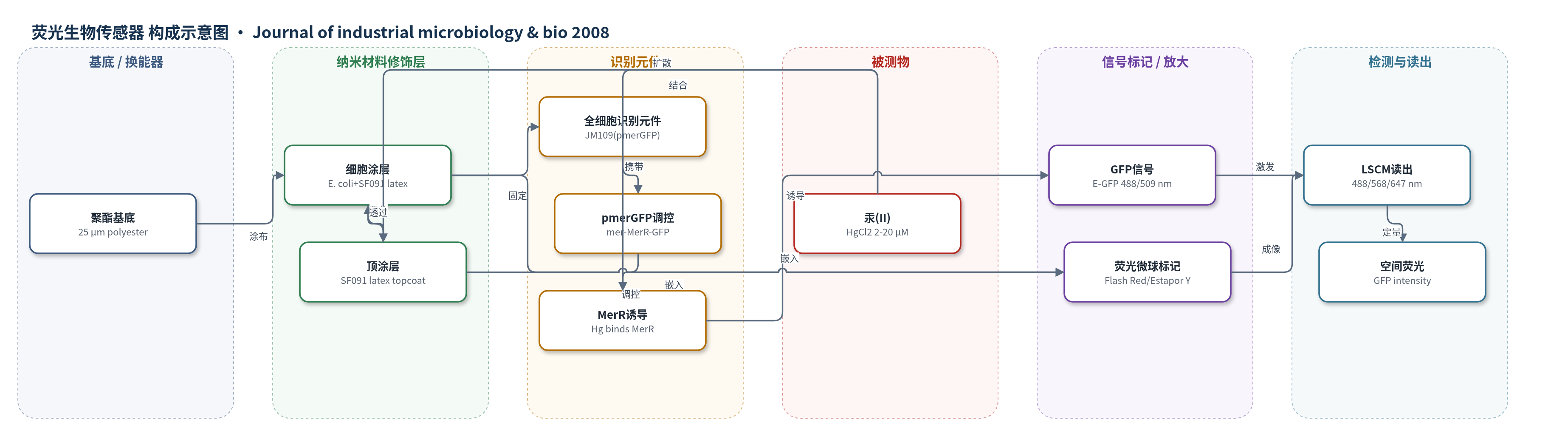
点击图片查看大图 · 依据论文自动绘制
传感器类型
荧光生物传感器
检测对象
汞(II)(mercury(II), Hg(II),以氯化汞 HgCl2 形式);样品基质:水相丙酮酸缓冲液(pyruvate buffer)
检测原理
该传感器以固定化大肠杆菌 JM109(pmerGFP) 为识别元件。Hg(II) 从水相扩散穿过纳米多孔乳胶顶涂层和细胞涂层,进入细胞后与 MerR 阻遏蛋白结合,改变 mer 启动子/操纵子的转录状态,诱导 gfp 基因表达。E-GFP 在氧存在下折叠并发出绿色荧光(488 nm 激发、509 nm 发射)。激光扫描共聚焦显微镜同时检测 GFP 和两种荧光微球,按 Z 轴逐层成像,从而获得涂层内 GFP 的空间分布。Hg(II) 浓度越高,MerR 诱导越强,GFP 荧光强度越高;由于 Hg(II) 在涂层中的扩散和与细胞表面结合,表达常从顶涂层–细胞涂层界面向底部呈梯度。
检测灵敏度
原文未报告 LOD、灵敏度斜率或相关系数;浓度范围: 2–20 μM HgCl2。
效应效果
在 2–20 μM HgCl2、21–37 °C、0–39 h 下,GFP 荧光随浓度、温度和时间增加;37 和 21 °C 下 2 到 20 μM 总荧光约增 2 倍,30 °C 约增 25%。37 °C 在 10、20 μM 表达显著更高。20 μM 诱导速率 88.3 RFU/h,10、5 μM 为 45.8、41.6,2、1 μM 为 20.4、11.3;前 25 h 速率高于后 15 h。顶涂层 10、21、37 μm 未显著影响表达,说明 Hg(II) 和氧扩散不受限。与 mer-lux 传感器相比,后者 100 nM、5 h 达 6×10^6 cpm,本 GFP 需 20 μM、24 h 达 650 RFU,Lux 更灵敏,但 GFP 适合原位空间监测。
传感器的构成
- 基底:25 μm 透明聚酯薄膜(3M polyester substrate),提供机械支撑与光学观察基底。
- 细胞涂层:E. coli JM109(pmerGFP)、SF091 乳胶(280 nm 丙烯酸/醋酸乙烯共聚物乳液)、甘油、Tween 20 和 Flash Red 荧光微球(0.19 μm,660/690 nm),固定全细胞识别元件并标记细胞层。
- 顶涂层:SF091 乳胶、甘油、Tween 20 和 Estapor Y 荧光微球(0.19 μm,555/570 nm),形成纳米多孔保护层并标记顶涂层。
- 识别元件:pmerGFP 质粒(mer 启动子/操纵子、merR 和 E-GFP)在大肠杆菌中,使 Hg(II) 诱导 GFP 表达。
- 信号标记物:E-GFP(488/509 nm),汞诱导后产生绿色荧光。
- 层标记微球:Flash Red 与 Estapor Y 荧光微球,用于 LSCM 区分细胞涂层与顶涂层。
- 孵育介质:丙酮酸缓冲液(5 mM pyruvate、34 mM Na phosphate、33 mM K phosphate、pH 6.8、0.091 mM (NH4)2SO4)及 HgCl2,维持细胞活性并提供诱导剂。
中文摘要
为优化作为生物传感器或生物催化剂的细胞涂层,需要测量固定化细胞中的基因表达。本研究构建质粒 pmerGFP,将绿色荧光蛋白(GFP)基因置于汞抗性操纵子调控序列的转录控制下,用于定量和定位乳胶基汞生物传感器中的基因表达。含该质粒的大肠杆菌 JM109 暴露于氯化汞后诱导合成 GFP,并可通过荧光定量。将 JM109(pmerGFP) 与 SF091 乳胶、Tween 20 和甘油混合,在聚酯基底上涂成约 20 μm 厚的纳米多孔粘附细胞涂层,再覆盖含乳胶、Tween 20 和甘油的纳米多孔顶涂层。不同荧光微球分别标记顶涂层和细胞涂层。暴露 Hg(II) 后,涂层内细胞诱导产生 GFP,并用激光扫描共聚焦显微镜(LSCM)在空间上定量表达。GFP 表达随汞浓度(2–20 μM)、温度(21–37 °C)和孵育时间(0–39 h)增加而升高;细胞涂层中 GFP 表达呈梯度,顶涂层–细胞涂层界面附近高于底部。顶涂层厚度未显著影响 GFP 表达,表明 Hg(II) 和氧通过顶涂层的扩散不构成限制。
英文摘要
Optimizing the reactivity of cell coatings developed as biosensors or biocatalysts requires measurements of gene expression in the immobilized cells. To quantify and localize gene expression within a latex-based mercury biosensor, a plasmid, pmerGFP, was constructed, which contains the green fluorescent protein (GFP) gene under transcriptional control of the mercury resistance operon regulatory sequences. When cells containing this plasmid were exposed to mercuric chloride, GFP synthesis was induced and could be quantified by fluorescence. E. coli strain JM109 (pmerGFP) was mixed with SF091 latex (Rohm & Haas), Tween 20, and glycerol, and coated as an approximate 20-microm thick nanoporous adhesive coating on a polyester substrate. The cell coat was overlaid with a nanoporous topcoat of latex, Tween 20, and glycerol. Different fluorescent microspheres were used to mark the topcoat and cell coat layers of the coating. Upon exposure to mercury(II), cells within the coating were induced to synthesize GFP, and laser scanning confocal microscopy was used to quantify expression spatially within the cell coat. GFP expression in the coatings increased with increasing mercury concentration (2-20 microM), temperature (21-37 degrees C), and time of incubation (0-39 h). There was a gradient of GFP expression through the cell coat with expression higher near the topcoat-cell coat interface relative to the bottom of the cell coat. The topcoat thickness did not significantly affect GFP expression indicating that diffusion of mercury(II) and oxygen through the topcoat was not limiting.