组成图示
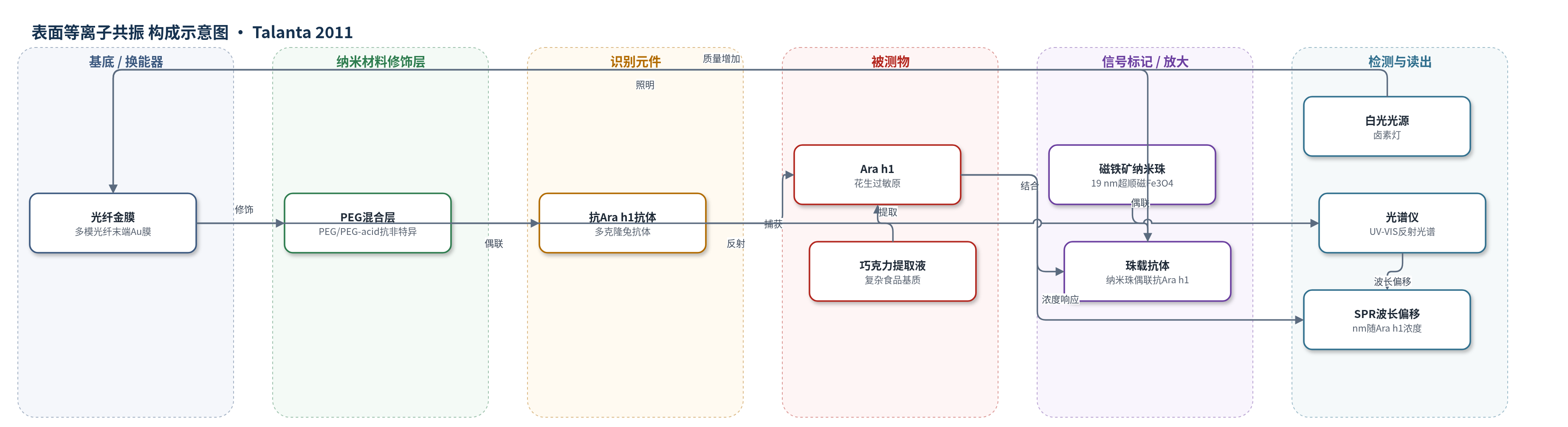
点击图片查看大图 · 依据论文自动绘制
传感器类型
表面等离子共振(SPR)生物传感器
检测对象
花生主要过敏原 Ara h1(Arachis hypogaea 1);样品基质:巧克力糖果棒提取上清(复杂食品基质)
检测原理
该传感器基于光纤表面等离子共振(SPR)与纳米珠二级标记放大。多模光纤末端金膜在白光照射下激发表面等离子体,反射光谱出现SPR凹陷;金表面先修饰PEG/PEG-acid混合层,再经EDC/NHS固定多克隆抗Ara h1抗体。样品中的Ara h1与表面抗体结合后,加入表面偶联抗Ara h1抗体的超顺磁磁铁矿纳米珠,形成夹心结构。纳米珠具有较大质量(约11280 kDa),使界面质量显著增加,导致SPR共振波长发生红移。波长偏移量随Ara h1浓度增加而增大,在0.1–2 μg/mL范围内呈线性。超顺磁纳米珠便于处理,无微流道设计避免堵塞,酸处理可快速再生。
检测灵敏度
LOD: 0.09 μg/mL(无标记 9 μg/mL,二抗夹心 0.21 μg/mL);线性范围: 0.1–2 μg/mL;灵敏度: 1 nm shift per 28.6 ng/cm2;分辨率: 5×10⁻⁵ RIU
效应效果
该传感器在巧克力复杂基质中抗干扰良好:提取前加标、提取后加标和缓冲液加标的ELISA剂量响应曲线几乎完全重叠,说明提取过程未造成灵敏度损失。抗体对Ara h1特异,仅在高浓度下对Ara h2/Ara h6有有限交叉反应。PEG混合层和垂直表面抑制纳米珠非特异结合,负对照表明非特异相互作用受控。SPR光纤可用pH 1.7甘氨酸缓冲液2 min再生,单根重复使用35次无明显灵敏度损失。与商品化多克隆ELISA相比,两者Ara h1浓度良好相关;SPR线性范围0.1–2 μg/mL大于ELISA(ELISA在1 μg/mL饱和),检测约20 min,比最快ELISA约45 min快两倍。无微流道避免堵塞和冲洗,组件可微型化,适合食品快速检测。
传感器的构成
- 基底/换能器:多模光纤末端金膜(Au),激发表面等离子体并反射光谱
- 抗非特异封闭层:聚乙二醇/聚乙二醇酸混合自组装单分子层(PEG/PEG-acid,4:1 v/v,Poly-pure),降低非特异吸附
- 识别元件:多克隆兔抗Ara h1抗体(anti-Ara h1 IgG),经EDC/NHS偶联至PEG羧基,捕获花生过敏原
- 信号标记/放大:超顺磁磁铁矿纳米珠(Fe3O4,约19 nm),经N-(三甲氧基硅基丙基)乙二胺三乙酸三钠盐/3-[双(2-羟乙基)氨基]丙基三乙氧基硅烷硅烷化,并用EDC/NHS偶联多克隆抗Ara h1抗体,作为二级标记放大SPR信号
- 封闭/储存:1% BSA PBS-T(BSA/PBS-T),封闭非特异位点并储存传感器
- 再生液:pH 1.7甘氨酸缓冲液(glycine buffer),酸处理再生金表面抗体层
中文摘要
本文首次报道一种纳米珠增强光纤表面等离子共振(SPR)生物传感器,并将其用于复杂食品基质中花生过敏原的快速准确检测。作者以巧克力糖果棒为样品,比较了无标记检测、二抗夹心检测和纳米珠增强免疫检测三种策略。虽然无标记检测操作简便,但功能化纳米珠可显著改善光纤SPR检测限。以超顺磁磁铁矿纳米颗粒作为二级标记后,Ara h1的SPR检测限由9 μg/mL提高至0.09 μg/mL,提升约两个数量级。纳米颗粒的超顺磁性便于样品处理,SPR光纤可经酸处理再生,单根光纤最多可重复使用35次而无明显灵敏度损失。与商品化多克隆ELISA试剂盒相比,两种方法测得的Ara h1浓度具有良好相关性;SPR光纤检测速度约为最快ELISA方案的两倍,且无需微流道,可避免堵塞和耗时冲洗。该传感器线性动态范围为0.1–2 μg/mL,明显大于ELISA基准。
英文摘要
This paper is the first report of a fiber optic SPR biosensor with nanobead signal enhancement. We evaluated the system with a bioassay for the fast and accurate detection of peanut allergens in complex food matrices. Three approaches of an immunoassay to detect Ara h1 peanut allergens in chocolate candy bars were compared; a label-free assay, a secondary antibody sandwich assay and a nanobead enhanced assay. Although label-free detection is the most convenient, our results illustrate that functionalized nanobeads can offer a refined solution to improve the fiber SPR detection limit. By applying magnetite nanoparticles as a secondary label, the detection limit of the SPR bioassay for Ara h1 was improved by two orders of magnitude from 9 to 0.09 μg/mL. The super paramagnetic character of the nanoparticles ensured easy handling. The SPR fibers could be regenerated easily and one fiber could be reused for up to 35 times without loss of sensitivity. The results were benchmarked against a commercially available polyclonal ELISA kit. An excellent correlation was found between the Ara h1 concentrations obtained with the ELISA and the concentrations measured with the SPR fiber assay. In addition, with the SPR fiber we could measure the samples twice as fast as compared to the fastest ELISA protocol. Since the dipstick fiber has no need for microchannels that can become clogged, time consuming rinsing step could be avoided. The linear dynamic range of the presented sensor was between 0.1 and 2 μg/mL, which is considerably larger than the ELISA benchmark.