组成图示
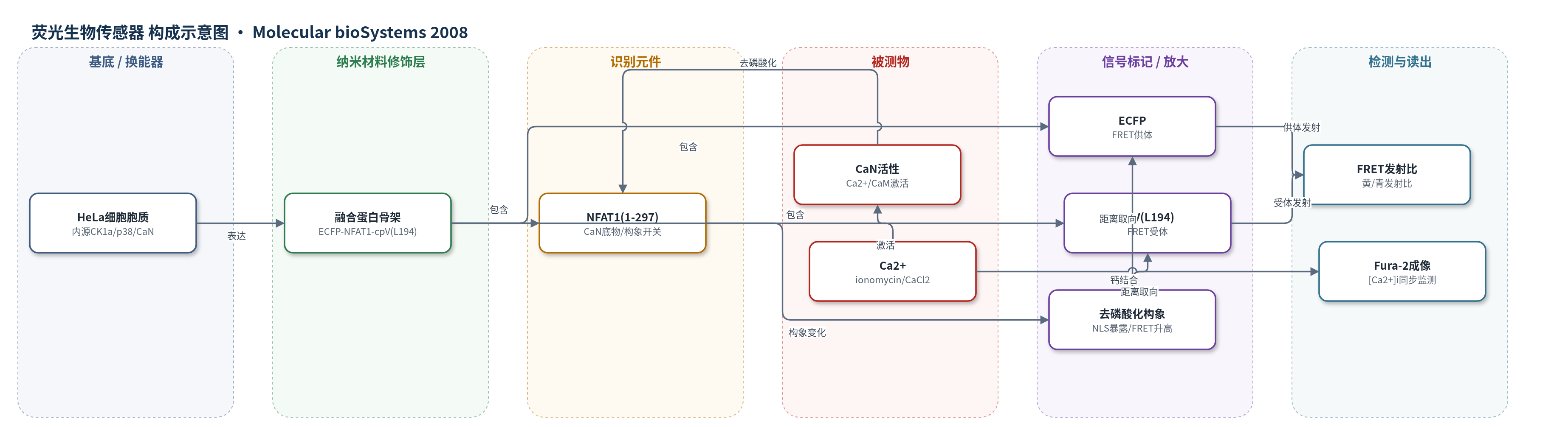
点击图片查看大图 · 依据论文自动绘制
传感器类型
荧光生物传感器
检测对象
钙调磷酸酶活性(calcineurin activity, CaN activity);样品基质:HeLa活细胞胞质
检测原理
静息HeLa细胞中,NFAT1(1-297)调控域被内源激酶CK1a和p38磷酸化,磷酸基团与核定位信号NLS中的正电荷残基发生静电作用,掩盖NLS,使ECFP与cpV(L194)保持较大距离或不利偶极取向,FRET效率较低。加入ionomycin/CaCl2后,细胞内Ca2+升高,Ca2+结合钙调蛋白CaM并激活CaN;CaN特异性去磷酸化NFAT1(1-297),暴露NLS并引起构象变化,改变ECFP-cpV(L194)的相对距离和取向,使FRET效率升高,黄/青发射比增加。发射比随CaN去磷酸化程度增加而升高,CsA预处理可完全阻断该变化,表明信号来源于CaN活性。
检测灵敏度
原文未报告LOD、线性范围、灵敏度斜率或相关系数。
效应效果
CaNAR1在HeLa细胞中主要分布于胞质。加入1 μM ionomycin和5 mM CaCl2后,黄/青发射比12 min内升至最大6.3±1.5%,t1/2为3.5 min,平台维持数分钟;撤除ionomycin后可回到静息水平。10 μM CsA预处理10 min完全消除FRET变化,显示CaN特异性。Western blot显示未诱导CaNAR1迁移约125 kDa,诱导后迁移率增加,CsA处理无变化,证明FRET变化与去磷酸化相关。Fura-2同步成像显示Ca2+峰值约3.5 min,与CaNAR1半最大响应同步。作者认为该遗传编码传感器可用于活细胞CaN活性的时空监测。
传感器的构成
- 荧光供体:ECFP,融合于NFAT1(1-297) N端,作为FRET供体
- 识别/底物:NFAT1 N端调控域NFAT1(1-297),含SRR-1、SP2、SP3和NLS,作为CaN去磷酸化底物与构象开关
- 荧光受体:cpV(L194),融合于NFAT1(1-297) C端,作为FRET受体
- 细胞工作基质:HeLa细胞胞质,提供内源激酶CK1a、p38及Ca2+/CaM-CaN作用环境
- 信号读出:荧光显微镜发射比成像,黄/青发射比变化反映CaN活性
中文摘要
蛋白激酶和磷酸酶在细胞内形成复杂的信号网络,其活性需要在空间和时间上精确协调。为理解信号转导网络的调控机制,有必要在天然细胞环境中定义蛋白激酶和磷酸酶的时空动态。本文报道了一种遗传编码蛋白生物传感器的开发,用于特异性探测钙离子/钙调蛋白依赖性蛋白磷酸酶钙调磷酸酶(calcineurin, CaN)的活性。该报告分子采用磷酸酶活性依赖的分子开关,以活化T细胞核因子(NFAT)N端调控域作为钙调磷酸酶的特异性底物,夹在增强青色荧光蛋白(ECFP)和黄色荧光蛋白(cpV(L194))之间。利用该报告分子,钙调磷酸酶活性可通过去磷酸化诱导的荧光共振能量转移(FRET)增强进行监测,并可同时成像细胞内钙动态。该原型磷酸酶活性传感器的成功设计为研究磷酸酶的靶向定位和区室化奠定了基础。
英文摘要
Protein kinases and phosphatases are organized into complex intracellular signaling networks designed to coordinate their activities in both space and time. In order to better understand the molecular mechanisms underlying the regulation of signal transduction networks, it is important to define the spatiotemporal dynamics of both protein kinases and phosphatases within their endogenous environment. Herein, we report the development of a genetically-encoded protein biosensor designed to specifically probe the activity of the Ca2+/calmodulin-dependent protein phosphatase, calcineurin. Our reporter design utilizes a phosphatase activity-dependent molecular switch based on the N-terminal regulatory domain of the nuclear factor of activated T-cells as a specific substrate of calcineurin, sandwiched between cyan fluorescent protein and yellow fluorescent protein. Using this reporter, calcineurin activity can be monitored as dephosphorylation-induced increases in fluorescence resonance energy transfer and can be simultaneously imaged with intracellular calcium dynamics. The successful design of a prototype phosphatase activity sensor lays a foundation for studying targeting and compartmentation of phosphatases.