组成图示
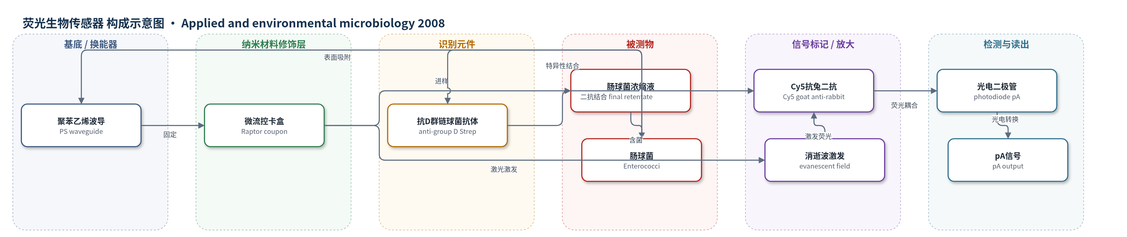
点击图片查看大图 · 依据论文自动绘制
传感器类型
荧光生物传感器
检测对象
肠球菌(Enterococci,含 Enterococcus faecalis 等);样品基质:佛罗里达坦帕湾娱乐海水/海滩水(recreational water/beach water),经100 L超滤浓缩最终浓缩液(final retentate)
检测原理
100 L海水经死端中空纤维超滤、超声、筛分和离心二次浓缩,使肠球菌浓度提高约4个数量级。最终浓缩液中的肠球菌吸附于聚苯乙烯波导表面,抗D群链球菌抗体特异性结合肠球菌,Cy5标记山羊抗兔二抗结合一抗。Raptor激光在波导中产生消逝波,激发表面100–1000 nm内Cy5荧光,部分发射耦合回波导并由光电二极管以pA读出。信号随肠球菌浓度增加而增强;以非目标抗M. tuberculosis抗体建立基线,SALOD大于0且高于阴性对照判阳性。方法主要依靠大体积浓缩和荧光免疫识别,无核酸或酶催化放大。
检测灵敏度
LOD: 1.0 × 10^5 CFU/ml(Fig. 3:间接 Raptor 法最低可检测肠球菌浓度);缓冲液阳性检测: ≥5.0 × 10^5 cells/ml;缓冲液检测范围: 5.0 × 10^4–5.0 × 10^8 cells/ml;r^2 = 0.94(P < 0.001);实际浓缩液SALOD与环境水浓度相关: R^2 = 0.59(P < 0.0001);逻辑回归 Nagelkerke R^2 = 0.61(P < 0.0001)。
效应效果
该法缓冲液中对粪肠球菌≥5.0×10^5 cells/ml阳性,平均SALOD 20,r^2=0.94。ELISA中抗D群链球菌抗体对多种肠球菌1.0×10^5–1.0×10^8 cells/ml S/N>2.0,1.0×10^8时S/N 14.8–18.1。12份现场海水最终浓缩液较环境水提高至少4个数量级,浓缩倍数1,000–约177,000倍,回收率4%–708%、平均251%。环境水超过>10^5 CFU/100 ml时SALOD 11–100,低于限值时SALOD<0;逻辑回归Nagelkerke R^2=0.61。全流程2.5 h,较EPA 1600的24 h和qPCR 3–4 h更快,处理100 L水样,可用于海滩快速预警。
传感器的构成
- 换能基底:聚苯乙烯波导(polystyrene waveguide, PS)作为消逝波光纤传感表面,肠球菌直接吸附
- 微流控载体:Raptor波导卡盒(coupon,含4根波导、光学胶固定、样品孔)用于进样与激光激发
- 识别元件:亲和纯化的兔抗D群链球菌多克隆IgG(anti-group D Streptococcus antibody)特异性结合肠球菌
- 信号标记物:Cy5标记山羊抗兔IgG(Cy5-labeled goat anti-rabbit secondary antibody)结合一抗并产生荧光
- 封闭/缓冲液:blocking buffer(1% BSA和酪蛋白于PBS)用于抗体孵育,降低非特异结合
- 基线对照抗体:兔抗M. tuberculosis抗体(anti-M. tuberculosis antibody)用于建立非目标基线
中文摘要
为快速监测娱乐水体的粪便污染,本研究建立了中空纤维超滤浓缩与生物传感器检测联用的肠球菌快速检测方法。在佛罗里达坦帕湾上湾采集12份100 L海水样品,现场采用死端中空纤维超滤浓缩,随后对初始浓缩液进行超声处理、微米级筛分以去除干扰颗粒,并离心二次浓缩;同时采集瞬时水样。采用EPA方法1600测定各阶段肠球菌浓度,用于计算回收率和浓缩倍数。初始浓缩液中肠球菌浓度较环境水提高约100倍,最终浓缩液提高约4个数量级。将最终浓缩液吸附到聚苯乙烯波导上,用Raptor微流控消逝波光纤生物传感器进行免疫分析。当环境水肠球菌浓度超过单样品监管限值(>10^5 CFU/100 ml)时可检出。该联用流程仅需2.5 h,而标准方法需24 h,证明可用于现场快速水质监测。
英文摘要
Monitoring recreational waters for fecal contamination by standard methodologies involves culturing indicator bacteria, such as fecal coliforms and enterococci. Delayed reporting of microbial water quality parameters increases the likelihood of public exposure to pathogens of fecal origin, making the development of rapid methods important for public health protection. A rapid assay for enterococci was developed using a combined ultrafiltration-biosensor procedure. Twelve 100-liter water samples were collected from upper Tampa Bay over a 9-month period. The samples were collected on site by dead-end hollow-fiber ultrafiltration. Postfiltration processing of the initial retentates included sonication and micrometer-level sieve passage to remove interfering particles. Centrifugation was utilized for secondary concentration. Grab samples were collected simultaneously with the ultrafiltered samples. Concentrations of enterococci in all grab and ultrafiltration samples were determined by the standard method (EPA method 1600) for calculation of recovery efficiencies and concentration factors. Levels of enterococci increased twofold in initial retentates and by 4 orders of magnitude in final retentates over ambient concentrations. An aliquot of each final retentate was adsorbed onto polystyrene waveguides for immunoassay analysis of enterococci with a microfluidic fiber optic biosensor, the Raptor. Enterococci were detected when concentrations in the ambient water exceeded the regulatory standard for a single sample (> or =105 CFU/100 ml). The combined ultrafiltration-biosensor procedure required 2.5 h for detection compared to 24 for the standard method. This study demonstrated that enterococci can be detected rapidly using on-site ultrafiltration, secondary concentration, and biosensor analysis.