组成图示
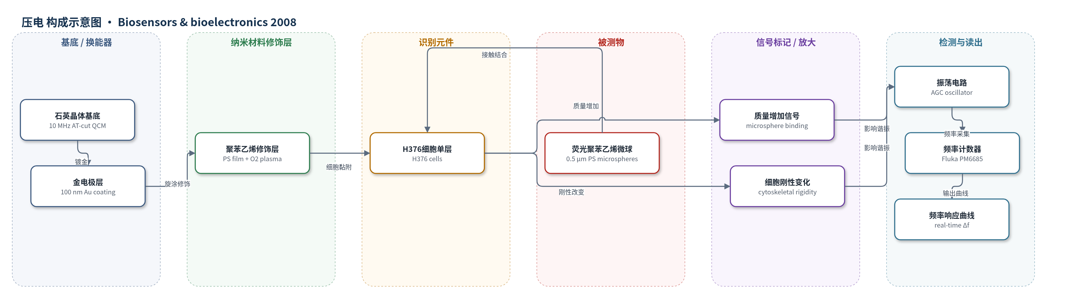
点击图片查看大图 · 依据论文自动绘制
传感器类型
压电(QCM)生物传感器
检测对象
人源口腔上皮细胞 H376 与荧光聚苯乙烯微球(fluorescent polystyrene microspheres, PS)的相互作用;样品基质:DMEM/F12 培养基(含 15 mM HEPES,37 °C、5% CO2 平衡)
检测原理
QCM 基于压电石英晶体谐振频率对表面质量与黏弹性敏感。H376 细胞单层作为识别与信号元件,微球到达后与细胞表面结合并被内吞,引起传感器界面质量增加,按 Sauerbrey 关系使谐振频率下降;同时微球刺激细胞膜并触发细胞骨架重塑,改变细胞刚性与黏弹性,可产生正频率偏移及后续负偏移。流动池中连续进样,振荡电路维持晶体振荡,频率计数器以 1 s 间隔记录 Δf,从而实时反映细胞-微球结合、内吞和细胞刚性变化。该体系未使用酶或核酸放大,信号增强主要来自细胞群体响应与内吞过程。
检测灵敏度
未报告
效应效果
无细胞传感器对重复微球进样保持响应,三颗晶体间频率变化 CV 为 15.0%。细胞传感器出现正频率峰值(35.49、46.96、60.98 Hz)及后续负偏移(-50.06、-38.89、-28.59 Hz)。Hoechst/PI 显示暴露组与对照组细胞数量无显著差异,说明剪切振荡和血清缺失不损害细胞。实验排除温度、压力、黏度/密度与渗透压伪影。作者认为可用于口腔流动条件下细胞-微粒相互作用研究、制剂筛选和细胞生物学研究。
传感器的构成
- 换能器基底:10 MHz AT-cut 石英晶体(QCM),表面镀 100 nm Au,作为压电谐振与电极基底
- 表面修饰层:聚苯乙烯(PS)薄膜(0.5% w/v 甲苯溶液旋涂)并经 O2 等离子处理,形成亲水细胞黏附表面
- 识别/信号元件:人源口腔上皮细胞 H376 单层,结合并内吞微球,将细胞-微粒相互作用转换为频率变化
- 流动池/进样系统:flow cell、PEEK HPLC 管路、Harvard 注射泵和 Rheodyne 9725 MBB 进样阀,维持 37 °C 流动并注入微球
- 读出系统:带 AGC 的表面安装振荡电路与 Fluka PM6685 频率计数器,实时记录谐振频率
中文摘要
石英晶体微天平(QCM)谐振传感技术近年来被用于监测细胞黏附与特异性配体相互作用,推动了 QCM 生物传感器的发展。本研究将人源口腔上皮细胞 H376 培养在石英传感器表面,并利用 QCM 技术原位研究其对微球的响应。结果表明,该新型生物传感器能够在模拟口腔流动条件下实时跟踪细胞与微球的相互作用。微球诱导的独特频率曲线被归因于质量增加和细胞刚性改变两个阶段。显微证据表明,这些独特频率响应部分来自细胞表面与微球之间的结合;此外,微球负载还引发细胞摄取过程,该过程通过影响细胞骨架刚性,也可在频率响应中被检测到。
英文摘要
Recent applications of quartz crystal resonant sensor technology to monitor cell adhesion and specific ligand interaction processes has triggered the development of a new category of quartz crystal microbalance (QCM) based biosensors. In this study human oral epithelial cells (H376) were cultured on quartz sensors and their response to microspheres investigated in situ using the QCM technique. The results demonstrated that this novel biosensor was able to follow cell-microsphere interactions in real-time and under conditions of flow as would occur in the oral cavity. Unique frequency profiles generated in response to the microspheres were postulated to be due to phases of mass addition and altered cellular rigidity. Supporting microscopic evidence demonstrated that the unique frequency responses obtained to these interactions were in part due to binding between the cell surface and the microspheres. Furthermore, a cellular uptake process, in response to microsphere loading was identified and this, by influencing the rigidity of the cellular cytoskeleton, was also detectable through the frequency responses obtained.