组成图示
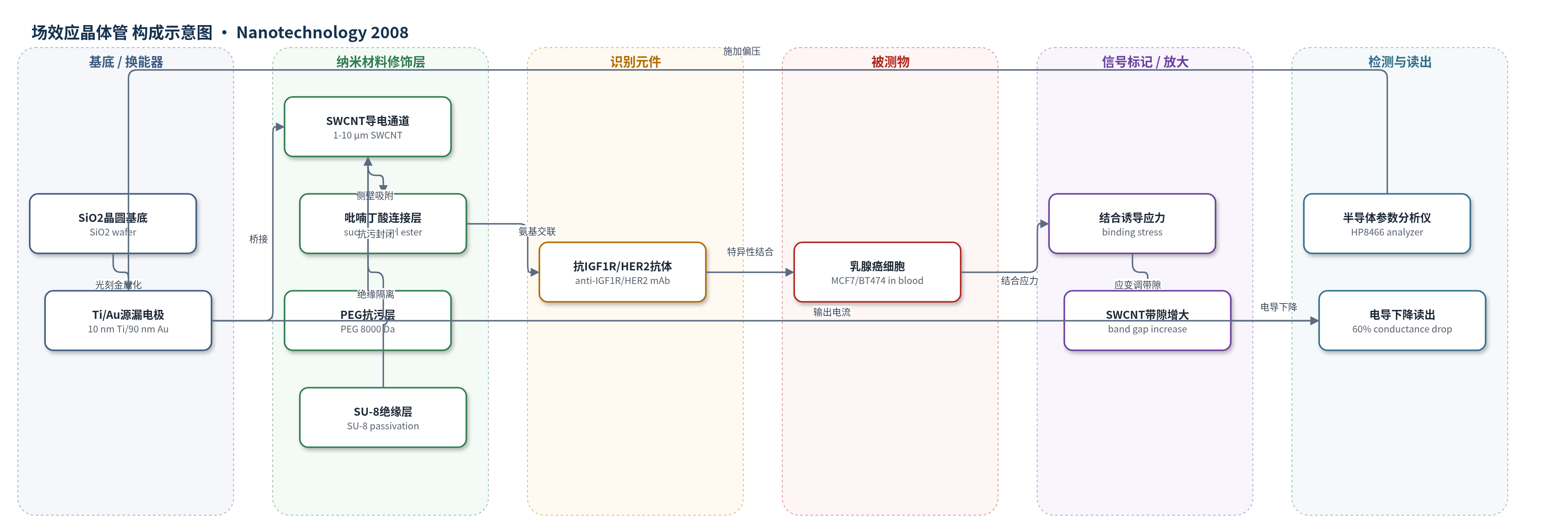
点击图片查看大图 · 依据论文自动绘制
传感器类型
场效应晶体管(FET)生物传感器
检测对象
人乳腺癌细胞(MCF7、BT474,circulating breast cancer cells);样品基质:新鲜人血(fresh human blood)
检测原理
该传感器为无标记单壁碳纳米管场效应晶体管(SWCNT FET)。SWCNT侧壁通过succinimidyl 1-pyrenebutanoate固定抗IGF1R或抗HER2单克隆抗体。当新鲜人血中MCF7或BT474乳腺癌细胞到达电极间隙时,细胞表面过表达的IGF1R或Her2受体与纳米管上的抗体发生多价特异性结合。多个抗原-抗体结合事件释放自由能,在受电极约束的SWCNT表面产生拉伸或扭转应力/应变,改变C-C键长度和纳米管带隙;对p型SWCNT而言,带隙增大使源漏电导下降。光学观察提示单个细胞结合即可引起可测电导变化,因此信号随目标细胞是否结合呈开关式变化,而非依赖外源标记或酶催化放大。
检测灵敏度
单细胞灵敏度;电导下降:60%;对照电导下降:<5%
效应效果
该器件在新鲜人血中直接检测,1 μl血样使电流从22.5 μA降至约15 μA后稳定,表明无明显生物污损,归因于PEG抗污层和1 μm电极间隙。特异性方面,anti-IGF1R/MCF7和anti-HER2/BT474配对电导下降约60%,非特异性抗体或MCF10A对照下降小于5%(结果中部分对照小于10%)。重复性方面,实验至少重复三次,三个不同anti-IGF1R器件对MCF7响应相似,器件间差异小于10%。光学显微镜显示单个细胞结合即可引起电导变化,支持单细胞灵敏度。作者认为该无标记、低成本、可阵列化器件可用于手持式直接血样检测循环肿瘤细胞、蛋白或DNA。
传感器的构成
- 基底/换能器电极:SiO2晶圆(SiO2 wafer)为基底,光刻图案化10 nm Ti/90 nm Au源漏电极,电极间距约1 μm,提供导电通道与电学读出。
- 纳米材料修饰层:单壁碳纳米管(SWCNT)桥接电极,长度1–10 μm、直径约15 nm,作为导电通道和应力敏感换能层。
- 连接/固定层:succinimidyl 1-pyrenebutanoate通过π-堆积吸附于SWCNT侧壁,其琥珀酰亚胺酯与抗体氨基交联,用于固定抗体。
- 识别元件:抗IGF1R小鼠单克隆抗体(anti-IGF1R mAb, Ab-3)和抗HER2单克隆抗体(anti-HER2 mAb, OP.39),分别识别乳腺癌细胞表面IGF1R和Her2受体。
- 封闭/抗污层:聚乙二醇(PEG,平均分子量8000 Da)覆盖未占据SWCNT侧壁,减少非特异性生物分子吸附。
- 绝缘隔离层:SU-8聚合物覆盖器件周围区域,隔离非目标区域并降低噪声。
中文摘要
近期研究表明,纳米电子器件可在受体蛋白与表面功能化抗体结合时检测电学性质变化。本文首次报道一种单壁碳纳米管场效应晶体管(SWCNT FET)阵列,分别功能化IGF1R特异性和Her2特异性抗体,可在新鲜人血中灵敏、选择性地检测完整活MCF7和BT474人乳腺癌细胞。两种细胞系均过表达IGF1R和Her2,但水平不同。功能化相应抗体的单根或小束SWCNT器件与BT474或MCF7细胞在2 μl血滴中作用时,电导下降约60%;非特异性抗体或MCF10A对照细胞电导下降小于5%。作者推测,多个细胞-抗体结合事件释放的自由能在纳米管表面产生应力,使带隙增大、电导下降。由于该自由能变化、应力和电导变化具有抗原-抗体特异性,可作为循环癌细胞分子传感指纹。光学观察显示单个细胞结合单个SWCNT FET即可引起电导变化,表明该纳米级肿瘤检测器具有单细胞灵敏度,并可在新鲜血滴中工作。
英文摘要
Recent reports have shown that nanoscale electronic devices can be used to detect a change in electrical properties when receptor proteins bind to their corresponding antibodies functionalized on the surface of the device, in extracts from as few as ten lysed tumor cells. We hypothesized that nanotube-antibody devices could sensitively and specifically detect entire live cancer cells. We report for the first time a single nanotube field effect transistor array, functionalized with IGF1R-specific and Her2-specific antibodies, which exhibits highly sensitive and selective sensing of live, intact MCF7 and BT474 human breast cancer cells in human blood. Those two cell lines both overexpress IGF1R and Her2, at different levels. Single or small bundle of nanotube devices that were functionalized with IGF1R-specific or Her2-specific antibodies showed 60% decreases in conductivity upon interaction with BT474 or MCF7 breast cancer cells in two µl drops of blood. Control experiments with non-specific antibodies or with MCF10A control breast cells produced a less than 5% decrease in electrical conductivity, illustrating the high sensitivity for whole cell binding by these single nanotube-antibody devices. We postulate that the free energy change due to multiple simultaneous cell-antibody binding events exerted stress along the nanotube surface, decreasing its electrical conductivity due to an increase in band gap. Because the free energy change upon cell-antibody binding, the stress exerted on the nanotube, and the change in conductivity are specific to a specific antigen-antibody interaction; these properties might be used as a fingerprint for the molecular sensing of circulating cancer cells. From optical microscopy observations during sensing, it appears that the binding of a single cell to a single nanotube field effect transistor produced the change in electrical conductivity. Thus we report a nanoscale oncometer with single cell sensitivity with a diameter 1000 times smaller than a cancer cell that functions in a drop of fresh blood.