组成图示
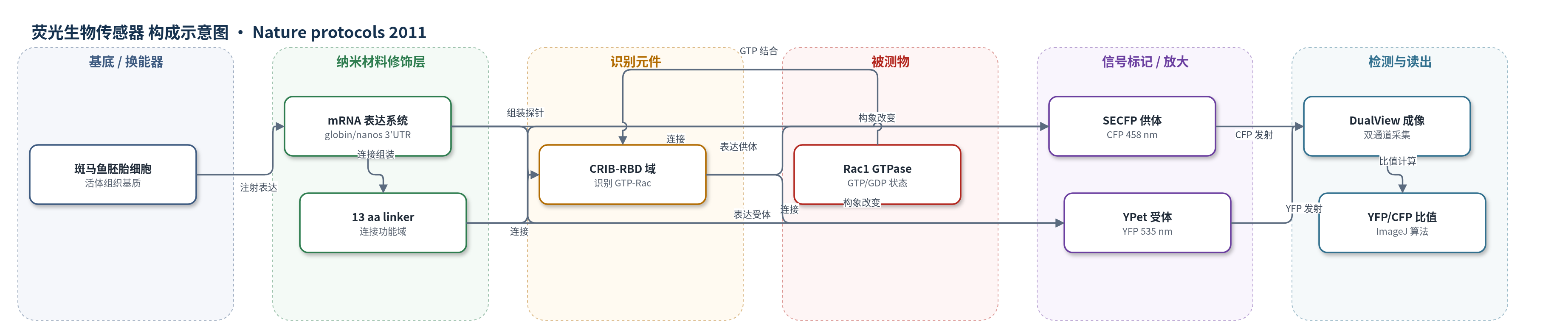
点击图片查看大图 · 依据论文自动绘制
传感器类型
荧光生物传感器
检测对象
Rac1 GTPase 活性(Rac1 GTPase activity);样品基质:活体斑马鱼胚胎细胞/组织(somatic cells、primordial germ cells)
检测原理
单链分子内 FRET 探针由 CFP/SECFP 供体、YPet/YFP 受体、CRIB-RBD 传感器域和 Rac1 配体域通过 linker 连接。当细胞内 Rac1 处于 GTP 结合激活态时,CRIB-RBD 与 Rac1 相互作用,使供体与受体距离缩短,FRET 效率升高。在 458 nm CFP 激发下,CFP 发射降低、YFP 发射升高,YFP/CFP 比值随 Rac 活性增加而增加。DualView 同时采集 CFP 和 YFP 通道,ImageJ 进行背景校正、图像配准和比值计算,从而在活体胚胎中实时、亚细胞分辨率读出蛋白激活状态。该方法无酶催化放大,主要依赖 FRET 距离敏感性和荧光蛋白量子产率,并用 nonlinked/linked 及 active/inactive 对照校准信号范围。
检测灵敏度
原文未报告 LOD、线性范围、灵敏度斜率或 R^2。
效应效果
该方案强调活体胚胎中低表达、低光照和严格对照。nonlinked 与 linked 对照分别定义无 FRET 和最大 FRET 的 YFP/CFP 范围;RacV12-FRET、RacFRET+Rac1N17 和 Tiam1 DH-PH 用于建立活性范围,示例中 RacFRET 工作范围约为 2–3.5。优化后的 RacFRET-NoCT 亚细胞定位与 mCherry-Rac1 一致,表达 24 h 后胚胎发育正常。完整流程需 2–3 d,可检测迁移 PGC 前缘 Rac 激活等空间模式。未报告 RSD、回收率或与 ELISA/HPLC/qPCR 的定量对比;主要限制是组织透明度和背景/配准伪影。
传感器的构成
- 样品基质/表达平台:活体斑马鱼胚胎细胞或组织(somatic cells、primordial germ cells),通过 mRNA 注射表达 FRET 探针,作为活体成像基质。
- 探针表达元件:体外转录 mRNA(mMessage mMachine 合成),含 Xenopus globin 3′UTR 或 nanos 3′UTR,用于全局或谱系特异性表达。
- 供体荧光蛋白:SECFP/CFP(donor),在 458 nm 激发下发射 CFP 通道,作为 FRET 供体。
- 受体荧光蛋白:YPet/YFP(acceptor),接收 FRET 能量并发射 YFP 通道,作为 FRET 受体。
- 识别元件:CRIB-RBD 传感器域(sensor domain),识别并结合 GTP 结合态 Rac1,引起构象变化。
- 配体/定位模块:Rac1 结构域(ligand domain,含 C-terminal targeting domain,优化为 NoCT/NoNLS),与 CRIB-RBD 相互作用并决定亚细胞定位。
- 连接元件:13 aa linker,连接 CFP/SECFP、CRIB-RBD、Rac1 和 YPet/YFP,维持单链探针构象。
中文摘要
基于荧光共振能量转移(FRET)的分子生物传感器是研究活细胞中蛋白活性的重要工具,但因其固有的细胞复杂性和三维成像挑战,很少用于完整生物体。本文提供在早期发育中使用单链分子内 FRET 生物传感器的实验方案,给出胚胎 FRET 比值成像的通用流程,包括数据采集条件和比值图像生成算法。作者以 pRaichu RacFRET 生物传感器为例,展示如何针对活体斑马鱼胚胎进行特定探针的适配与优化。方案涵盖探针 mRNA 注入胚胎、培养至目标发育阶段、去卵黄膜与固定、荧光显微镜采集 CFP/YFP 双通道图像,以及使用 ImageJ 进行背景校正、图像配准和 YFP/CFP 比值图生成。优化后的完整流程,包括探针引入、成像和数据分析,需要 2–3 天。
英文摘要
Fluorescence resonance energy transfer (FRET)-based molecular biosensors serve as important tools for studying protein activity in live cells and have been widely used for this purpose over the past decade. However, FRET biosensors are rarely used in the context of the live organism because of the inherent high cellular complexity and imaging challenges associated with the three-dimensional environment. Here we provide a protocol for using single-chain intramolecular FRET-based biosensors in early development. We provide a general protocol for FRET ratio imaging in embryos, including the data-acquisition conditions and the algorithm for ratio image generation. We then use the pRaichu RacFRET biosensor to exemplify the adaptation and optimization of a particular biosensor for use in live zebrafish embryos. Once an optimized biosensor is available, the complete procedure, including introduction of the probes into embryos, imaging and data analysis, requires 2-3 d.