组成图示
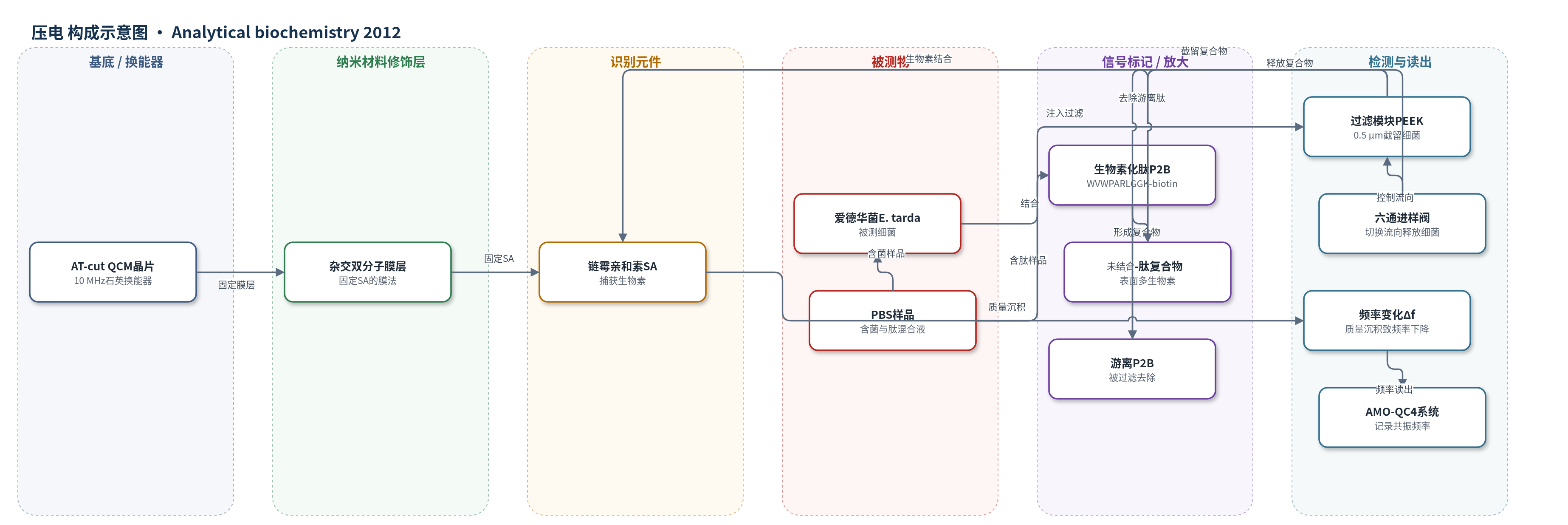
点击图片查看大图 · 依据论文自动绘制
传感器类型
压电(QCM)生物传感器
检测对象
爱德华菌(Edwardsiella tarda, E. tarda);样品基质:PBS 稀释细菌悬液
检测原理
本方法基于噬菌体肽库筛选得到的 E. tarda 结合肽 P2。将生物素化肽 P2B 与细菌混合后,P2B 特异性结合到 E. tarda 表面,使单个菌体携带多个生物素;随后通过 0.5 μm PEEK 过滤模块截留细菌-肽复合物,并洗去游离 P2B。反转六通阀流向后,复合物被输送至链霉亲和素(SA)包被的 QCM 芯片。细菌表面多个生物素与 SA 发生多重强结合,使菌体以较刚性方式固定在石英表面,质量沉积导致 AT 切石英晶振频率下降。菌量越高,沉积质量越大,频率变化越大,从而在 8×10^2–8×10^6 细胞范围内实现浓度依赖检测。荧光偏振法则利用小分子荧光肽 P2F 结合大细菌后旋转减慢,使偏振值升高。
检测灵敏度
QCM: LOD: 800 cells;线性范围: 8 × 10^2 to 8 × 10^6 cells。FP: 线性范围: 5.2 × 10^3 to 2.1 × 10^5 cells。Plate: 线性范围: 10^5 to 10^8 cells;LOD: 7 × 10^5 cells。
效应效果
选择性方面,P2 肽对 E. tarda 显示高亲和,而对大肠杆菌(E. coli)和肠耶尔森菌(Y. enterocolitica)无显著 FP 变化;过滤法中 P2B-SA-AP 复合物也仅对 E. tarda 保留明显活性。QCM 检测中,单独 E. tarda 或单独 P2B 均无显著频率变化,只有二者混合后出现浓度依赖频率下降,说明检测依赖肽介导的结合。QCM 下限为 800 个细胞,约 0.5 ng;FP 在 2.1×10^5 细胞时达 310 mP,可能接近上限。作者认为 FP 适合较清洁样品的高通量分析,QCM 过滤法因分离步骤可去除污染物,更适合复杂样品。文中未报告 RSD、稳定性、实际样品回收率或与 ELISA 的定量对比。
传感器的构成
- 换能器基底:AT-cut 10 MHz QCM 石英晶片(8×8 mm),提供压电振荡频率读出
- 固定层:杂交双分子膜(hybrid bilayer membrane)法制备的 SA 固定层,用于在 QCM 表面固定链霉亲和素
- 识别/捕获元件:链霉亲和素(SA),与细菌表面生物素多重结合
- 识别配体:生物素化七肽 P2B(WVWPARLGGK-biotin),特异性结合 E. tarda 并在菌体表面引入生物素
- 信号标记物:生物素(biotin),作为亲和标记介导细菌与 SA 芯片结合
- 过滤分离模块:0.5 μm PEEK 烧结片与六通进样阀,截留细菌-肽复合物并去除游离 P2B
- 样品介质:PBS 缓冲液,用于细菌稀释、肽孵育、洗涤和输送
中文摘要
本研究从噬菌体展示七肽库中筛选出可结合爱德华菌(Edwardsiella tarda, E. tarda)的肽配体,并分别以荧光素和生物素标记该肽,建立两种敏感检测方法。首先,荧光标记肽用于荧光偏振(FP)检测。由于 FP 灵敏度在荧光团连接小分子并结合大分子靶标时最高,小肽-荧光素探针适合检测细菌。该 FP 方法可在 5.2×10^3 至 2.1×10^5 个细胞范围内检测 E. tarda。其次,作者设计了连接过滤模块的石英晶体微天平(QCM)生物传感器。将 E. tarda 与生物素化肽的混合液注入过滤模块后,细菌-肽复合物被 0.5 μm 滤膜截留,未结合肽被去除;随后反转流向,将复合物输送至链霉亲和素(SA)包被的 QCM 芯片。含生物素化肽和 E. tarda 的样品在 8×10^2 至 8×10^6 个细胞范围内产生浓度依赖的频率变化。两种方法有望促进其他细菌结合肽检测方法的开发。
英文摘要
In this study, we identified a peptide ligand for Edwardsiella tarda from a phage peptide library and tested two approaches for sensitive detection of the bacteria with the peptide labeled with fluorescein or biotin. At first, the fluorescent peptide was proved to be advantageous in the fluorescence polarization (FP) assay because sensitivity of the assay is maximized when a fluorophore is linked to a small molecule. The FP assay using the fluorescent peptide enabled detection of E. tarda in a range from 5.2×10(3) to 2.1×10(5) cells. Second, we devised a new assay method using a quartz crystal microbalance (QCM) biosensor connected to a filter module. When a mixture of E. tarda and the biotinylated peptide was injected into the filter module, the E. tarda-peptide complex was separated from the unbound peptide by a filter and detected with a streptavidin-coated QCM sensor chip. On injection of samples containing the biotinylated peptide and E. tarda, concentration-dependent frequency change was observed in a range from 8×10(2) to 8×10(6) cells. The two approaches are expected to facilitate development of assay methods using other bacteria-binding peptides.