组成图示
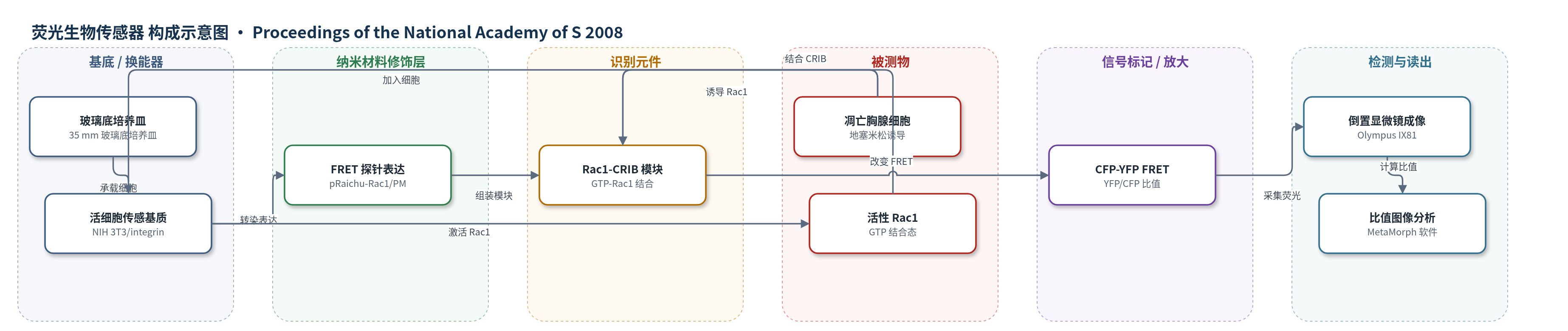
点击图片查看大图 · 依据论文自动绘制
传感器类型
荧光生物传感器
检测对象
活性 Rac1(active Rac1 / GTP-bound Rac1);样品基质:活吞噬细胞/成纤维细胞(NIH 3T3/integrin、Swiss 3T3/integrin、BAM3),在凋亡胸腺细胞刺激下
检测原理
Raichu-Rac1/PM 探针由 CFP、Rac1、Pak CRIB 基序、YFP 和 K-Ras C 端组成。当 Rac1 结合 GTP 时,CRIB 与 Rac1 结合,使 YFP 靠近 CFP,CFP 激发后能量转移至 YFP,YFP/CFP 比值升高;当 Rac1 结合 GDP 时,CRIB 脱离,CFP 与 YFP 距离增大,FRET 效率降低。凋亡胸腺细胞暴露磷脂酰丝氨酸,经 MFG-E8 桥接与吞噬细胞 αvβ3 整合素结合,诱导整合素聚集并激活/招募 Rac1 至吞噬杯。Rac1 激活驱动肌动蛋白聚合形成吞噬杯;吞噬杯闭合时 Rac1 失活,FRET 信号下降。该过程无需外源标记,通过活细胞荧光比值实时反映 Rac1 活性变化。
检测灵敏度
原文未报告 LOD、线性范围、灵敏度斜率或相关系数。
效应效果
该 FRET 探针可在活细胞中实时成像 Rac1 激活,信号对 Rac1 具有特异性:GTPase 失活突变 Raichu-Rac1G12V/PM 和 Raichu-RhoA/PM 的 FRET 效率变化很小。实验中 40/44 个凋亡胸腺细胞在片足区域被吞噬;在 35 个吞噬至少 4 个凋亡细胞的吞噬细胞中,86% 的细胞出现超过 2 个凋亡细胞从同一门户进入。组成型活性 Rac1Q61L 使吞噬杯闭合显著延迟,吞噬效率降低,Rac1 在吞噬杯上的停留时间平均延长 4 倍。BAM3 巨噬细胞中肌动蛋白斑组装约 2 min,解聚、吞噬杯闭合和 Rac1 下调在 1 min 内同步发生。作者认为该传感器适用于解析吞噬凋亡细胞过程中 Rac1 的时空调控。
传感器的构成
- 成像基底:35 mm 玻璃底培养皿(glass-base dish),承载细胞并支持倒置显微镜成像
- 细胞传感基质:NIH 3T3/integrin、Swiss 3T3/integrin 或 BAM3 细胞,作为活细胞表达平台;NIH/Swiss 3T3/integrin 表达 αvβ3 整合素
- 荧光供体:CFP(cyan fluorescent protein),433 nm 激发、475 nm 发射,提供 FRET 供体信号
- 荧光受体:YFP(yellow fluorescent protein),527 nm 发射,接收 FRET 能量并输出受体荧光
- 识别元件:Rac1 与 Pak CRIB 基序(CRIB)组成的 Raichu 模块,结合 GTP-Rac1 后改变 CFP-YFP 距离
- 膜定位元件:K-Ras C 端(Raichu-Rac1/PM)或 Rac1 C 端(Raichu-Rac1),将探针锚定至质膜或细胞内
- 信号读出:Olympus IX81 倒置显微镜、CoolSNAP HQ CCD 相机与 MetaMorph 软件,采集 CFP/YFP 并计算 YFP/CFP 比值
中文摘要
吞噬凋亡细胞需要吞噬细胞协调激活调控肌动蛋白动力学的 Rho 家族 GTP 酶。本研究利用 FRET 生物传感器实时可视化吞噬凋亡细胞过程中 Rac1 的时空激活。结果显示,凋亡细胞通常被吞噬细胞的片足吞噬,而片足区域 Rac1 处于激活状态;同一片足部位常连续吞噬多个凋亡细胞,提示存在凋亡细胞进入的门户。在该位置,激活的 Rac1 被招募并形成由肌动蛋白斑组成的吞噬杯。当吞噬杯闭合时,Rac1 活性下调,肌动蛋白斑迅速解体。组成型活性 Rac1 在吞噬杯上的停留时间明显长于野生型 Rac1,表达组成型活性 Rac1 的细胞中吞噬杯闭合显著延迟,导致吞噬效率降低。结果表明,激活的 Rac1 对 F-肌动蛋白组装是必要的,而吞噬杯闭合则需要 Rac1 失活。
英文摘要
The engulfment of apoptotic cells requires phagocytes to coordinately activate Rho family GTPases that regulate actin dynamics. Here, we used a FRET biosensor to visualize the spatiotemporal activation of Rac1 during engulfment of apoptotic cells. We report that apoptotic cells were usually engulfed by the phagocytes' lamellipodia, where Rac1 was activated. Often, apoptotic cells were engulfed successively at the same lamellipodial site, suggesting the presence of portals for apoptotic cells. At this location, the activated Rac1 was recruited to form phagocytic cups that were comprised of actin patches. When the phagocytic cup was closed, Rac1 was down-regulated, and the actin patches were abruptly broken down. The constitutively active Rac1 remained at phagocytic cup for a longer period than the wild-type Rac1, and the closure of the phagocytic cup was significantly delayed in cells expressing a constitutive active form of Rac1, resulting in inefficient engulfment. These results indicate that activated Rac1 is necessary to assemble F-actin, but closing the phagocytic cup requires Rac1 to be deactivated.