组成图示
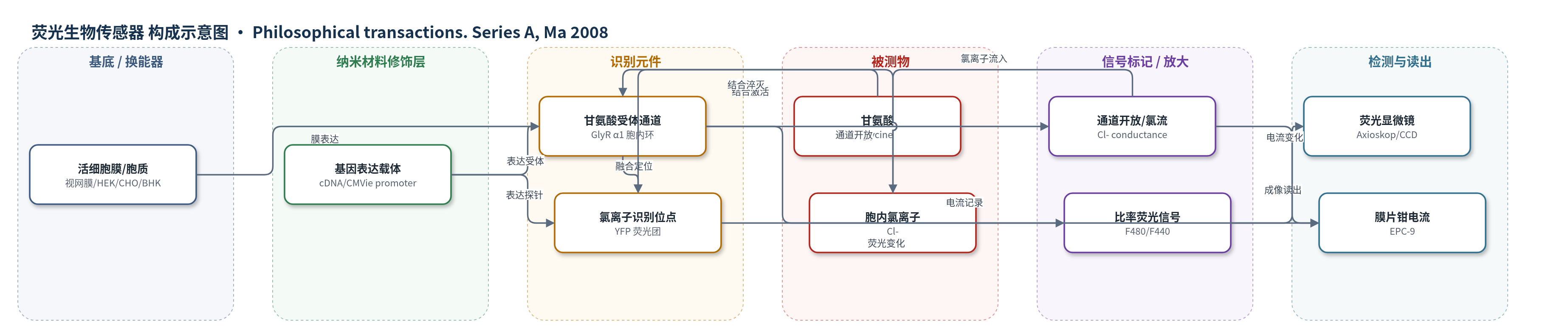
点击图片查看大图 · 依据论文自动绘制
传感器类型
荧光生物传感器
检测对象
胞内氯离子(intracellular chloride, [Cl]i)、甘氨酸(glycine)/甘氨酸受体通道活性(GlyR activity);样品基质:大鼠视网膜切片、HEK-293、CHO-K1、BHK-21 细胞
检测原理
Cl-Sensor 由 CFP 与 YFP 融合而成,YFP 荧光团与 Cl- 结合后发生荧光淬灭和激发光谱位移,CFP 对 Cl- 不敏感,作为内参。通过 440 nm 与 480 nm 交替激发并计算 F480/F440 比率,可比率读出 [Cl]i;Kapp≈30 mM,接近生理范围。高 K+ 去极化使 Cl- 内流,[Cl]i 升高,比率随之改变。BioSensor-GlyR 将 Cl-Sensor 插入 GlyR α1 胞内长环,甘氨酸结合受体外域后通道开放,Cl- 沿电化学梯度流入,通道附近胞质 Cl- 局部升高,使 Cl-Sensor 荧光变化。荧光变化幅度与甘氨酸诱导电流近似线性,因此可直接光谱监测受体门控通道活性。
检测灵敏度
原文未报告 LOD、线性范围、灵敏度斜率或相关系数;报告 Cl-Sensor Kapp≈30 mM;GlyR 甘氨酸 EC50:野生型 30–210 mM,BioSensor-GlyR 70–300 mM。
效应效果
Cl-Sensor 经体内电穿孔在大鼠视网膜表达,表达可维持超过一个月,光感受器中 P6 [Cl]i 为 51±2 mM,P16 为 31±1 mM,P21 为 36±1 mM;40 mM KCl 去极化使 [Cl]i 升高 43±3 mM,低 Cl- 溶液中仅 4±1 mM,显示 Cl- 依赖性和可逆性。BioSensor-GlyR 保留野生型 GlyR 的动力学、甘氨酸 EC50(野生型 30–210 mM,BioSensor-GlyR 70–300 mM)和 Cl- 选择性,反转电位接近 ECl。甘氨酸诱导电流与荧光变化近似线性,约 300 pA 电流即可产生可检测荧光。作者认为其可用于药物筛选、生理/病理 Cl- 监测及受体通道活性光谱监测。
传感器的构成
- 基因模板/表达载体:Cl-Sensor cDNA 或 BioSensor-GlyR cDNA,含 CMVie 启动子,用于在细胞内表达荧光探针或嵌合受体
- 细胞基底/微环境:大鼠视网膜切片、HEK-293、CHO-K1、BHK-21 细胞,提供膜、胞质及 Cl- 微环境
- 膜定位识别元件:人源 α1 亚基甘氨酸受体(GlyR)胞内长环,结合甘氨酸并介导 Cl- 通道开放,同时锚定 Cl-Sensor
- 离子识别/荧光换能元件:CFP–YFP 融合蛋白 Cl-Sensor,YFP 荧光团结合 Cl- 后激发光谱改变,CFP 作为参考
- 信号标记物:Cl-Sensor 的 CFP 与 YFP 荧光团,产生 440/480 nm 激发比率(F480/F440)变化
- 递送/转染系统:Lipofectamine 2000、Magnetofection CombiMag 或 in vivo 电穿孔,将 cDNA 导入细胞
中文摘要
基因编码探针已成为非侵入监测离子、蛋白分布及细胞组分迁移与形成的有力工具。本文报道两种分子探针的功能表达,用于非侵入荧光监测胞内氯离子浓度([Cl]i)和甘氨酸受体(GlyR)通道功能。第一种为近期开发的蓝荧光蛋白–黄荧光蛋白(CFP–YFP)融合探针 Cl-Sensor,对 Cl- 具有较高敏感性(Kapp≈30 mM)。作者通过体内电穿孔将其表达于视网膜细胞,并分析去极化及出生后三周内 [Cl]i 的变化。40 mM KCl 处理使 [Cl]i 升高约 40 mM;P6 大鼠视网膜光感受器平均 [Cl]i 约 50 mM,P16 和 P21 约 30–35 mM。第二种构象 BioSensor-GlyR 是将 Cl-Sensor 插入 GlyR 胞内结构域的通道蛋白,是首个用于光谱监测受体门控通道功能的分子探针。此类探针可用于药物筛选,并在不同生理和病理条件下监测 Cl- 及异源系统与神经元中 GlyR 活性。
英文摘要
Genetically encoded probes have become powerful tools for non-invasive monitoring of ions, distributions of proteins and the migration and formation of cellular components. We describe the functional expression of two molecular probes for non-invasive fluorescent monitoring of intracellular Cl ([Cl]i) and the functioning of glycine receptor (GlyR) channels. The first probe is a recently developed cyan fluorescent protein-yellow fluorescent protein-based construct, termed Cl-Sensor, with relatively high sensitivity to Cl (Kapp approximately 30 mM). In this study, we describe its expression in retina cells using in vivo electroporation and analyse changes in [Cl]i at depolarization and during the first three weeks of post-natal development. An application of 40 mM K+ causes an elevation in [Cl]i of approximately 40 mM. In photoreceptors from retina slices of a 6-day-old rat (P6 rat), the mean [Cl]i is approximately 50 mM, and for P16 and P21 rats it is approximately 30-35 mM. The second construct, termed BioSensor-GlyR, is a GlyR channel with Cl-Sensor incorporated into the cytoplasmic domain. This is the first molecular probe for spectroscopic monitoring of the functioning of receptor-operated channels. These types of probes offer a means of screening pharmacological agents and monitoring Cl under different physiological and pathological conditions and permit spectroscopic monitoring of the activity of GlyRs expressed in heterologous systems and neurons.