组成图示
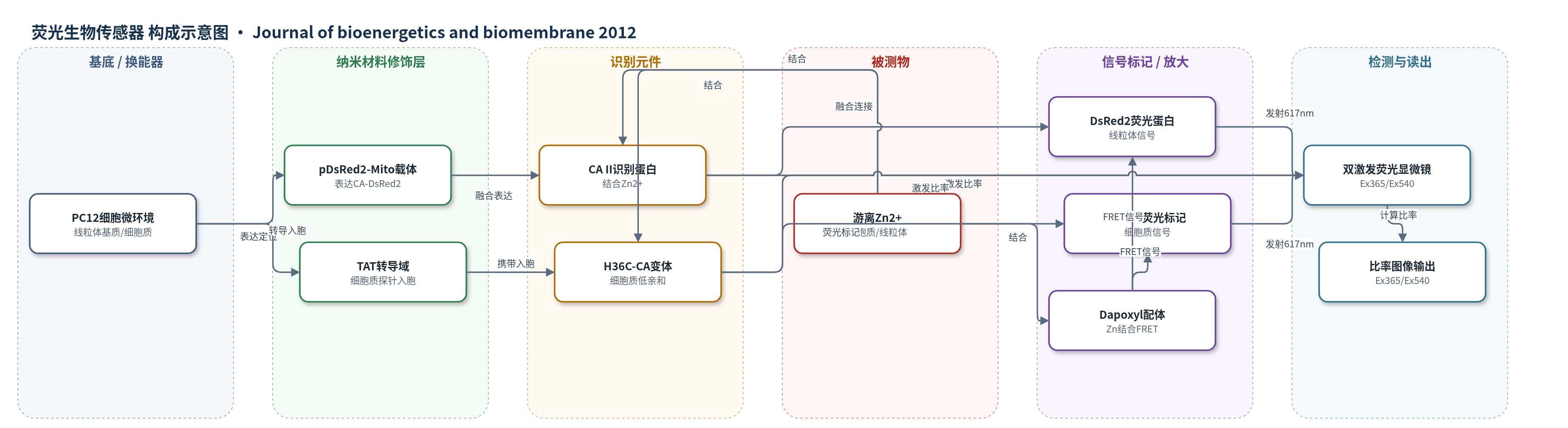
点击图片查看大图 · 依据论文自动绘制
传感器类型
荧光生物传感器
检测对象
游离锌离子(free Zn2+);样品基质:PC12细胞细胞质与线粒体基质(体外培养细胞,OGD缺血/再灌注模型)
检测原理
该传感器以人碳酸酐酶II(CA II)为识别元件,其活性位点结合游离Zn2+后改变金属配位环境,进而改变荧光配体Dapoxyl sulfonamide与DsRed2或AF594之间的FRET效率。Zn2+浓度升高时,激发比率Ex365/Ex540发生可逆变化;通过预先在分离线粒体或缓冲液中建立的校准曲线,可将比率转换为pM级游离锌浓度。线粒体定位序列将CA-DsRed2靶向线粒体基质,TAT域帮助细胞质探针进入细胞。比率成像可抵消细胞厚度、探针装载量、光漂白和自发荧光影响,无需外源酶放大。
检测灵敏度
Kd: 0.15±0.05 pM;动态范围: 0.02 pM–2 pM
效应效果
该比率荧光传感器避免强度型探针受细胞厚度、装载量、光漂白和自发荧光干扰,长发射约617 nm降低背景。作者用MitoTracker Green、TOM20/COX IV抗体及FCCP酸化实验确认探针位于线粒体基质。3 h OGD模型中,24 h约50%细胞死亡;细胞质游离锌由对照约10 pM降至1 h的0.9 pM,24 h升至>100 pM;线粒体游离锌由约0.2 pM在1 h升至>10 pM,2 h恢复。统计n=70–90(细胞质)、35–45(线粒体),p<0.05或p<0.01。相比FluoZin-3(KD约15 nM、非比率),可活细胞pM级定量。
传感器的构成
- 换能微环境:PC12细胞线粒体基质与细胞质,作为Zn2+结合和荧光信号产生的生物微环境
- 识别元件:人碳酸酐酶II(CA II)及其H36C变体,特异性结合Zn2+并改变荧光激发特性
- 信号标记物:DsRed2荧光蛋白(线粒体传感器)或Alexa Fluor 594(AF594)标记的TAT-H36C-CA(细胞质传感器),提供约617 nm发射
- 荧光配体:Dapoxyl sulfonamide(DS),与Zn2+结合并参与FRET/激发比率信号
- 定位元件:线粒体定位序列(MLS,来自COX IV亚基VIII)和TAT蛋白转导域,分别靶向线粒体基质和介导细胞质探针入胞
- 表达载体:pDsRed2-Mito质粒,用于在PC12细胞中表达CA-DsRed2融合蛋白
- 读出装置:Nikon Eclipse TE300荧光显微镜、Cooke Sensicam QE冷却CCD相机与IPLab软件,采集Ex365/Ex540双激发图像并计算比率
中文摘要
锌离子在缺血性卒中、癫痫持续状态和创伤性脑损伤后的细胞毒性作用仍有争议,但可能源于线粒体功能障碍。本文报道了一种基于人碳酸酐酶II(CA II)变体的激发比率荧光生物传感器,并将其表达于线粒体基质,用于定量成像其中的游离锌水平。在PC12大鼠嗜铬细胞瘤细胞培养系中,作者测得线粒体基质平均游离锌浓度约为0.2 pM。随后在细胞氧葡萄糖剥夺(OGD)缺血/再灌注模型中,对细胞质和线粒体游离锌水平进行成像。结果显示,3 h OGD后1 h,线粒体锌水平显著升高,而细胞质锌水平下降;随后线粒体锌恢复至生理水平,细胞质锌在存活细胞中于24 h内逐渐升高。OGD后复氧期间线粒体锌升高可能参与体外和体内再灌注模型中的生物能量功能障碍与细胞死亡。
英文摘要
The role of zinc ion in cytotoxicity following ischemic stroke, prolonged status epilepticus, and traumatic brain injury remains controversial, but likely is the result of mitochondrial dysfunction. We describe an excitation ratiometric fluorescence biosensor based on human carbonic anhydrase II variants expressed in the mitochondrial matrix, permitting free zinc levels to be quantitatively imaged therein. We observed an average mitochondrial matrix free zinc concentration of 0.2 pM in the PC12 rat pheochromacytoma cell culture line. Cytoplasmic and mitochondrial free zinc levels were imaged in a cellular oxygen glucose deprivation (OGD) model of ischemia/reperfusion. We observed a significant increase in mitochondrial zinc 1 h following 3 h OGD, at a time point when cytosolic zinc levels were depressed. Following the increase, mitochondrial zinc levels returned to physiological levels, while cytosolic zinc increased gradually over a 24 h time period in viable cells. The increase in intramitochondrial zinc observed during reoxygenation after OGD may contribute to bioenergetic dysfunction and cell death that occurs with both in vitro and in vivo models of reperfusion.