组成图示
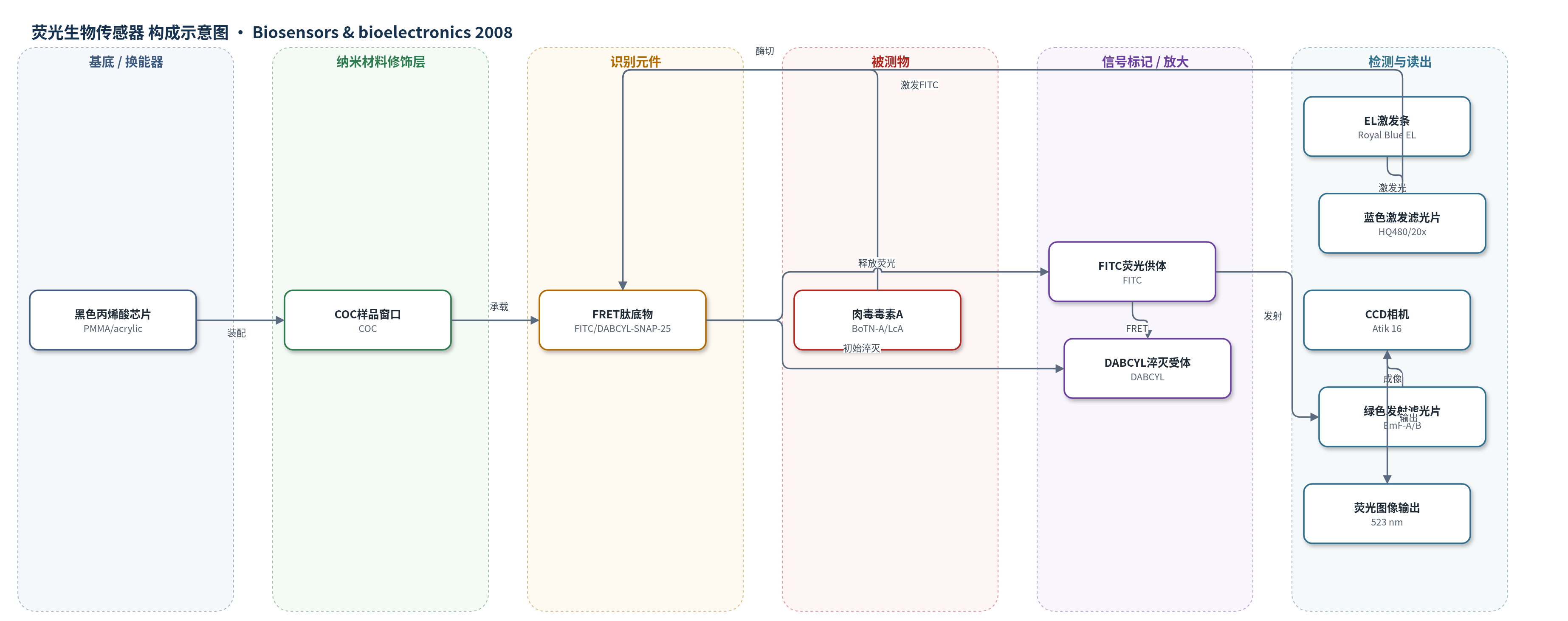
点击图片查看大图 · 依据论文自动绘制
传感器类型
荧光生物传感器
检测对象
肉毒神经毒素A(Botulinum neurotoxin A, BoTN-A)、肉毒神经毒素A轻链(LcA);样品基质为HEPES/Zn/BSA/DTT缓冲液(非实际临床/食品基质)
检测原理
检测基于FRET活性原理。FITC/DABCYL-SNAP-25肽底物中,FITC供体与DABCYL受体空间接近,发生Förster共振能量转移,FITC荧光被淬灭。当样品中存在BoTN-A或其轻链LcA时,毒素的蛋白酶活性切割SNAP-25肽序列,使供体与受体分离,FRET被破坏,FITC荧光在523 nm处增强。EL条经蓝色带通滤光片提供约494 nm空间激发光,激发FITC;绿色发射滤光片选择523 nm发射,冷却CCD相机成像,ImageJ分析平均强度。信号以信噪比(S/N)表示,随毒素浓度升高而增大,无需额外化学放大。
检测灵敏度
LOD: 0.625–1.25 nM(31–62 ng/ml)(LcA);LOD: 0.313 nM(45 ng/ml)(全毒素BoTN-A);LOD: 1.25 nM(62.5 ng/ml)(LcA);LOD: 0.625 nM(LcA,MicroAmp管);LOD: 0.625 nM(LcA,芯片内24 h);LOD: 2.5 nM(BoTN-A,2 h);LOD: 0.31 nM(BoTN-A,24 h)
效应效果
EL-CCD检测器与标准荧光酶标仪结果无显著差异(ANOVA P=0.843),LcA在2 h孵育后LOD为0.625–1.25 nM(31–62 ng/ml),全毒素BoTN-A经24 h孵育LOD为0.313 nM(45 ng/ml),与ELISA、侧流免疫检测等2–50 ng/ml水平相当,但低于磁珠电化学发光免疫检测的50 pg/ml。9孔间相对标准偏差为6.2%;COC、PC和玻璃基底无显著差异,Mylar因自荧光显著降低信噪比。PC顶盖使信号下降9.5%±1.8%。系统仅需12 µl样品,12 V DC供电,约825 mA,模块化便携,适合即时检测。
传感器的构成
- 样品芯片基底:黑色丙烯酸(PMMA/acrylic)9孔芯片,激光切割,承载样品并减少光散射与孔间串扰
- 样品池窗口:COC(cyclic olefin copolymer)底部窗口,高光学透明,承载12 µl样品
- 样品池顶盖:PC(polycarbonate)顶盖,可选密封样品,减少蒸发并允许孵育/储存
- 激发光源:Royal Blue EL条(电致发光半导体条),提供约494 nm蓝色空间激发光
- 激发滤光片:蓝色带通滤光片HQ480/20x,置于EL条上方,选择激发波长并抑制EL杂散绿光
- 识别/反应底物:FITC/DABCYL-SNAP-25 FRET肽底物,作为BoTN-A/LcA活性识别与信号报告元件
- 信号标记物:FITC供体荧光基团与DABCYL受体淬灭基团,切割后FITC在523 nm发射增强
- 封闭剂:BSA(bovine serum albumin)用于缓冲液和芯片预封闭,减少非特异结合
- 检测模块:冷却CCD相机(Atik 16/Sony ICX-429ALL)、绿色发射滤光片EmF-A/EmF-B及Pentax镜头,采集荧光图像
中文摘要
现有生物检测照明技术(激光、LED、钨灯等)多基于点照明,实现空间激发时需额外光学元件。本文提出一种基于电致发光(EL)半导体条的空间照明新方法,该条带可提供多种波长,简化生物传感器设计并消除额外光学部件。作者将EL激发与电荷耦合器件(CCD)荧光检测相结合,开发用于肉毒神经毒素A(BoTN-A)活性分析的简易便携检测器。系统包括检测模块(CCD相机与发射滤光片)和激发/样品模块(EL条、激发滤光片及9孔样品芯片)。研究采用FITC/DABCYL-SNAP-25肽底物的Förster共振能量转移(FRET)活性检测:BoTN-A或其轻链LcA切割底物后,FITC荧光发射增强。EL-CCD检测器测得的检出限与标准荧光酶标仪相近,LcA为0.625–1.25 nM(31–62 ng/ml),全毒素BoTN-A为0.313 nM(45 ng/ml)。据作者所知,这是首次将磷光体基EL条用于表面空间激发并结合CCD实现即时检测。
英文摘要
Current biodetection illumination technologies (laser, LED, tungsten lamp, etc.) are based on spot illumination with additional optics required when spatial excitation is required. Herein we describe a new approach of spatial illumination based on electroluminescence (EL) semiconductor strips available in several wavelengths, greatly simplifying the biosensor design by eliminating the need for additional optics. This work combines EL excitation with charge-coupled device (CCD) based detection (EL-CCD detector) of fluorescence for developing a simple portable detector for botulinum neurotoxin A (BoTN-A) activity analysis. A Förster Resonance Energy Transfer (FRET) activity assay for BoTN-A was used to both characterize and optimize the EL-CCD detector. The system consists of two modules: (1) the detection module which houses the CCD camera and emission filters, and (2) the excitation and sample module, containing the EL strip, the excitation filter and the 9-well sample chip. The FRET activity assay used in this study utilized a FITC/DABCYL-SNAP-25 peptide substrate in which cleavage of the substrate by BoTN-A, or its light chain derivative (LcA), produced an increase in fluorescence emission. EL-CCD detector measured limits of detection (LODs) were similar to those measured using a standard fluorescent plate reader with valves between 0.625 and 1.25 nM (31-62 ng/ml) for LcA and 0.313 nM (45 ng/ml) for the full toxin, BoTN-A. As far as the authors are aware this is the first demonstration of phosphor-based EL strips being used for the spatial illumination/excitation of a surface, coupled with CCD for point of care detection.