组成图示
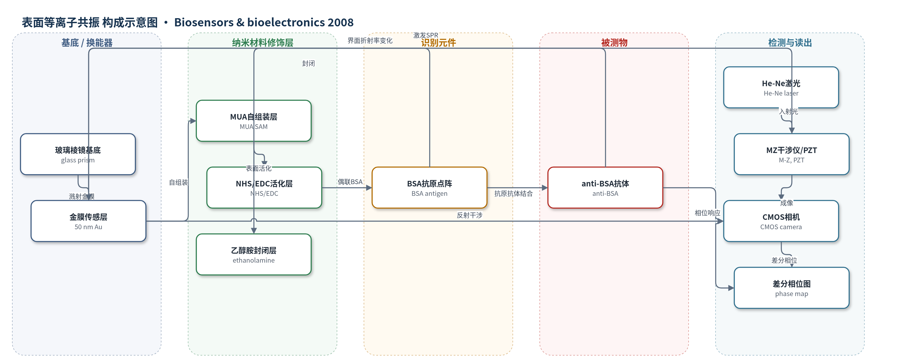
点击图片查看大图 · 依据论文自动绘制
传感器类型
表面等离子共振(SPR)生物传感器
检测对象
抗牛血清白蛋白抗体(anti-BSA antibody);样品基质:PBS 缓冲液中的蛋白微阵列样品
检测原理
传感器以金膜表面固定 BSA 抗原作为识别元件,anti-BSA 抗体从 PBS 样品中扩散至金表面并与 BSA 特异性结合。结合事件增加传感界面质量与局部折射率,使 Kretschmann 构型下 p 偏振光的 SPR 相位发生偏移;s 偏振光不受 SPR 激发,作为参考。Mach-Zehnder 干涉仪中 PZT 驱动参考镜做时域移相,CMOS 相机连续采集 p/s 偏振干涉图,FFT 提取各像素组相位,p-s 差分消除机械漂移等共模噪声。每个像素组对应一个传感点,形成二维相位图。抗体浓度越高,平衡相位变化越大,从而实现无标记实时定量监测。
检测灵敏度
LOD: 0.77 µg/ml(7.7 × 10^-4 mg/ml);系统分辨率: 8.8 × 10^-7 RIU
效应效果
盐水实验:0–12 wt% 对应折射率 1.3330–1.3541 RIU,最敏感区 1.3330–1.3347 RIU 相位变化 83.5°;8% 盐水 20 min 标准差饱和于 0.043°,分辨率 8.8×10^-7 RIU。BSA 点阵 A–D 浓度 2.5、1.3、0.6、0.3 mg/ml,E 为 PBS 对照;注入 0.025 mg/ml anti-BSA 后,A 点平衡相位变化为 D 点 1.7 倍、E 点 2.7 倍,B 点 33 min 标准差 0.88°、响应 28.6°,LOD 7.7×10^-4 mg/ml,与 Lee 等 10^-4 mg/ml 可比。对照点响应低,非特异结合不显著;共路差分相位抑制机械漂移,适用于高通量蛋白阵列。
传感器的构成
- 基底/换能器:玻璃棱镜(glass prism)与 50 nm 溅射金膜(Au),构成 Kretschmann 型 SPR 传感表面
- 自组装修饰层:11-巯基十一酸(MUA)自组装单层(SAM),在金表面提供羧基用于蛋白偶联
- 表面活化层:N-羟基琥珀酰亚胺(NHS)与 1-乙基-3-(3-二甲氨基丙基)碳二亚胺(EDC)混合液,活化羧基以共价固定 BSA
- 识别元件:牛血清白蛋白(BSA)抗原点阵,以不同浓度固定于金表面,用于捕获 anti-BSA 抗体
- 封闭层:乙醇胺(ethanolamine)溶液封闭未反应区域,降低非特异结合
- 样品装载/阵列结构:聚二甲基硅氧烷(PDMS)5×5 微孔阵列,每孔 5 µl,用于分装不同 BSA 样品并实现二维点阵
- 光学换能/读出:He-Ne 激光、Mach-Zehnder 干涉仪、压电移相镜(PZT)、Wollaston 棱镜与 CMOS 相机,采集 p/s 偏振干涉图并差分相位成像
中文摘要
本文报道了差分相位表面等离子共振(SPR)成像在二维蛋白质生物传感器阵列中的应用。与常规角度 SPR 技术相比,该相位成像方法在光学传感元件利用效率上具有明显优势:角度法中每个传感位点需要一维探测器阵列定位 SPR 角度凹陷,二维成像器件最多只能提供一维 SPR 生物传感器阵列;而相位敏感 SPR 在时域采集数据,干涉图中每个像素可代表一个传感位点,从而实现高密度二维生物传感器阵列。此外,差分相位方法通过消除共模干扰提高了检测分辨率。实验结果表明,系统分辨率为 8.8×10^-7 RIU。利用生物传感器阵列实现了不同浓度牛血清白蛋白(BSA)/抗 BSA 抗体结合相互作用的实时监测,检测限为 0.77 µg/ml。所报道的二维 SPR 生物传感器阵列为高通量蛋白质阵列分析提供了实时、无标记检测工具,有望应用于蛋白质治疗、药物筛选和临床诊断。
英文摘要
This paper reports the application of differential phase surface plasmon resonance (SPR) imaging in two-dimensional (2D) protein biosensor arrays. Our phase imaging approach offers a distinct advantage over the conventional angular SPR technique in terms of utilization efficiency of optical sensor elements in the imaging device. In the angular approach, each biosensor site in the biosensor array requires a linear array of optical detector elements to locate the SPR angular dip. The maximum biosensor density that a two-dimensional imaging device can offer is a one-dimensional SPR biosensor array. On the other hand, the phase-sensitive SPR approach captures data in the time domain instead of the spatial domain. It is possible that each pixel in the captured interferogram represents one sensor site, thus offering high-density two-dimensional biosensor arrays. In addition, our differential phase approach improves detection resolution through removing common-mode disturbances. Experimental results demonstrate a system resolution of 8.8 x 10(-7)RIU (refractive index unit). Real-time monitoring of bovine serum albumin (BSA)/anti-BSA binding interactions at various concentration levels was achieved using a biosensor array. The detection limit was 0.77 microg/ml. The reported two-dimensional SPR biosensor array offers a real-time and non-labeling detection tool for high-throughput protein array analysis. It may find promising applications in protein therapeutics, drug screening and clinical diagnostics.