组成图示
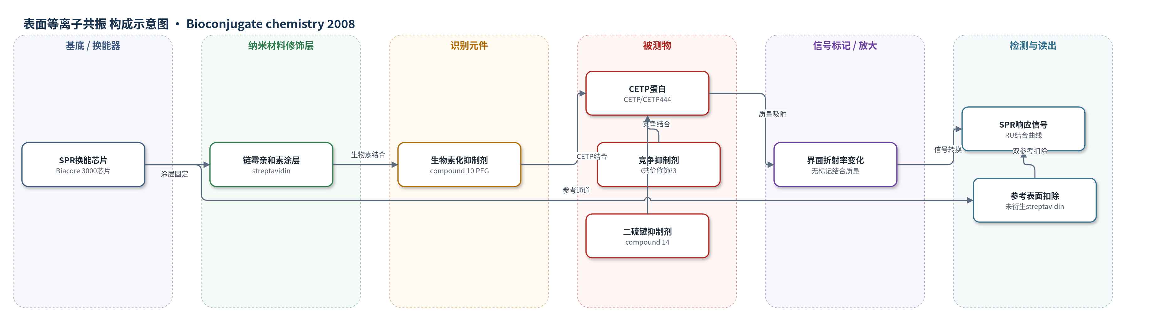
点击图片查看大图 · 依据论文自动绘制
传感器类型
表面等离子共振(SPR)生物传感器
检测对象
胆固醇酯转移蛋白(CETP, cholesteryl ester transfer protein);样品基质:重组CETP蛋白溶液(20 mM Tris pH 7.4、150 mM NaCl、0.005% Tween 20、1% DMSO),部分抑制实验使用90%人血浆。
检测原理
该SPR方法将生物素化torcetrapib类似物(compound 10)通过生物素-链霉亲和素相互作用固定在SPR芯片表面。CETP从溶液流过时,其N端脂质结合口袋与固定配体结合,使传感器界面附近质量与折射率增加,SPR角/响应单位(RU)随CETP浓度升高而增大并趋于饱和。结合动力学按1:1 Langmuir模型拟合,得到KD约0.3 µM。若CETP预先与游离torcetrapib类似物CP-532,623结合,或经二硫键抑制剂compound 14共价修饰Cys-13,则结合口袋被占据或阻断,表面结合信号下降。该方法为无标记、实时检测,未使用酶或荧光放大。
检测灵敏度
SPR动力学分析CETP浓度范围: 2.8 µM to 0.1 µM;KD ≈ 0.3 µM;ka = 22 000 ± 200 M^-1 s^-1;kd = 0.0064 ± 0.0001 s^-1。
效应效果
固定化配体长度决定CETP结合:2.1 nm compound 8无特异结合,琼脂糖柱容量约75 µg/mL;3.5 nm compound 9结合>98%,容量约1100 µg/mL;4.6 nm compound 10的SPR结合容量比compound 9高约25%。未衍生链霉亲和素表面结合<3%、容量<10 µg/mL。CETP与compound 10的结合可被等摩尔torcetrapib或CP-532,623竞争;二硫键抑制剂compound 14预孵育阻断结合并降低胆固醇酯转移活性>90%。质谱显示修饰质量增加约317 Da且可被DTT逆转,肽图定位Cys-13。作者认为SPR固定小分子配体可定位CETP抑制剂结合位点。
传感器的构成
- SPR换能芯片:Biacore 3000标准链霉亲和素涂层芯片,提供表面等离子共振检测与固定化平台
- 捕获层:链霉亲和素(streptavidin),特异性结合生物素化配体
- 固定配体/识别元件:生物素化torcetrapib类似物(compound 10,含4.6 nm PEG-biotin间隔臂),模拟抑制剂结合位点并捕获CETP
- 信号层:无标记CETP(野生型或CETP 444)结合,界面质量/折射率变化产生SPR信号
- 参考通道:未衍生链霉亲和素表面,用于双参考扣除缓冲液漂移
- 再生/清洗:0.1% SDS脉冲,去除结合CETP并恢复表面
- 运行缓冲液:20 mM Tris pH 7.4、150 mM NaCl、0.005% Tween 20、1% DMSO,维持CETP活性并降低非特异吸附
中文摘要
胆固醇酯转移蛋白(CETP)在不同类型血浆脂蛋白之间转移中性脂质,其抑制剂可提高血浆胆固醇中与高密度脂蛋白(HDL)结合的比例,正被开发用于心血管疾病的预防和治疗,但其分子机制尚未完全阐明。为研究抑制剂与CETP的相互作用,作者将torcetrapib类似物偶联到不同生物素终止间隔臂上,并在琼脂糖珠和表面等离子共振(SPR)生物传感器上监测CETP与链霉亲和素固定偶联物的结合。2.0 nm间隔臂结合较差,而3.5或4.6 nm聚乙二醇(PEG)间隔臂结合有效;4.6 nm偶联物用于后续SPR实验。可溶性抑制剂可阻断CETP与固定药物的结合,含二硫键共价抑制剂预孵育也产生阻断。为初步估计torcetrapib类似物的结合位点,作者用含二硫键试剂修饰CETP的Cys-13;质谱显示单个半分子共价结合,胃蛋白酶消化肽图确认Cys-13为修饰位点。修饰后的CETP不能结合SPR固定的torcetrapib类似物,表明torcetrapib结合位点位于蛋白N端附近的脂质结合口袋;晶体结构显示Cys-13巯基位于该口袋底部。
英文摘要
Cholesteryl ester transfer protein (CETP) transfers neutral lipids between different types of plasma lipoprotein. Inhibitors of CETP elevate the fraction of plasma cholesterol associated with high-density lipoproteins and are being developed as new agents for the prevention and treatment of cardiovascular disease. The molecular basis of their function is not yet fully understood. To aid in the study of inhibitor interactions with CETP, a torcetrapib-related compound was coupled to different biotin-terminated spacer groups, and the binding of CETP to the streptavidin-bound conjugates was monitored on agarose beads and in a surface plasmon resonance biosensor. CETP binding was poor with a 2.0 nm spacer arm, but efficient with polyethyleneglycol spacers of 3.5 or 4.6 nm. The conjugate based on a 4.6 nm spacer was used for further biosensor experiments. Soluble inhibitor blocked the binding of CETP to the immobilized drug, as did preincubation with a disulfide-containing covalent inhibitor. To provide a first estimate of the binding site for torcetrapib-like inhibitors, CETP was modified with a disulfide-containing agent that modifies Cys-13 of CETP. Mass spectrometry of the modified protein indicated that a single half-molecule of the disulfide was covalently bound to CETP, and peptide mapping after digestion with pepsin confirmed previous reports based on mutagenesis that Cys-13 was the site of modification. Modified CETP was unable to bind to the biosensor-mounted torcetrapib analog, indicating that the binding site on CETP for torcetrapib is in the lipid-binding pocket near the N-terminus of the protein. The crystal structure of CETP shows that the sulfhydryl group of Cys-13 resides at the bottom of this pocket.