组成图示
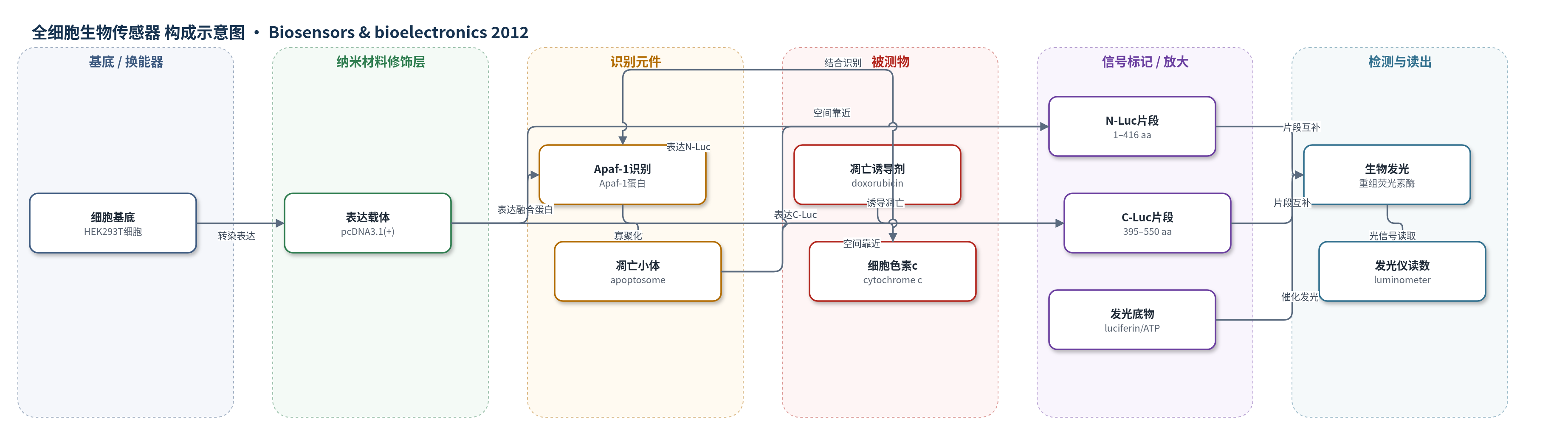
点击图片查看大图 · 依据论文自动绘制
传感器类型
全细胞生物传感器
检测对象
早期凋亡(apoptosis,细胞色素c释放/Apaf-1寡聚化/凋亡小体形成);样品基质:HEK293T 完整细胞、细胞裂解液
检测原理
阿霉素诱导线粒体途径凋亡,线粒体释放细胞色素c;细胞色素c结合Apaf-1的WD40结构域,诱导构象变化并暴露核苷酸结合位点,在(d)ATP结合与水解后,Apaf-1寡聚化形成轮状凋亡小体。N-Luc–Apaf-1与C-Luc–Apaf-1因凋亡小体组装而空间靠近,分裂荧光素酶片段互补重组,恢复部分荧光素酶活性。加入荧光素和ATP后,重组酶催化荧光素氧化发光,光强度随凋亡程度和细胞色素c释放量增加而升高。由于Apaf-1寡聚化具有协同性,时间响应呈S形,使该传感器可在caspase-3/7激活前动态监测早期凋亡。
检测灵敏度
原文未报告检出限、线性范围、灵敏度斜率或相关系数。
效应效果
该传感器在阿霉素诱导后4 h即产生显著生物发光,细胞裂解液信号较对照升高约15倍,完整细胞约6倍;10 h达最大,裂解液约155倍(讨论中亦称约150倍),随后逐渐下降。Apaf-1单体邻近和凋亡小体形成较caspase-3/7显著活性出现约早5 h,提示可检测早期凋亡。与FLICA荧光探针相比,生物发光背景更低、信噪比更高,避免细胞自荧光和探针非特异积累;与含DEVD位点的环化荧光素酶策略相比,无需长时间孵育等待酶切。作者认为其可用于化疗药物筛选、体内成像和临床前研究,并可构建稳定细胞系进行多细胞系时间/剂量依赖分析。
传感器的构成
- 细胞基底:HEK293T 人胚胎肾细胞,作为全细胞表达与凋亡响应平台
- 表达载体:pcDNA3.1(+) 质粒(CMV 启动子),携带 N-Luc–Apaf-1 与 C-Luc–Apaf-1 构建体
- 识别元件:Apaf-1 蛋白(CARD/NOD/WD40 结构域),结合细胞色素 c 并寡聚化形成凋亡小体
- 信号报告元件:分裂荧光素酶 N-Luc(1–416 aa)与 C-Luc(395–550 aa),片段互补后恢复生物发光
- 连接元件:(G4S)3 与 (G4S)4 柔性连接肽,连接荧光素酶片段与 Apaf-1 并促进正确折叠
- 诱导刺激物:阿霉素(doxorubicin),诱导线粒体途径凋亡及细胞色素 c 释放
- 发光底物:荧光素(luciferin)与 ATP,支持重组荧光素酶催化发光反应
- 信号读出:生物发光仪(luminometer,Berthold Detection System),检测光强度
中文摘要
本文报道一种基于Apaf-1寡聚化和凋亡小体形成、利用分裂荧光素酶策略检测早期凋亡的新型全细胞生物传感器。将荧光素酶N端片段(1–416 aa)和C端片段(395–550 aa)分别融合至Apaf-1 N端,构建N-Luc–Apaf-1和C-Luc–Apaf-1表达载体,共转染HEK293T细胞。用阿霉素诱导凋亡后,线粒体释放细胞色素c促使Apaf-1寡聚化形成凋亡小体,使两个荧光素酶片段空间靠近并部分重组,恢复生物发光活性。通过生物发光检测24 h内程序性细胞死亡,发现诱导后4 h信号较对照升高约15倍,10 h达最大约155倍;Apaf-1单体邻近和凋亡小体形成较caspase-3/7显著活性出现约早5 h。时间响应曲线呈S形,提示细胞色素c结合诱导Apaf-1协同寡聚化。该传感器可作为蛋白片段互补平台,用于评估潜在化疗药物及凋亡时间/剂量依赖研究。
英文摘要
We present here a novel whole-cell biosensor to detect early-stages of apoptosis based on Apaf-1 oligomerization and apoptosome formation using the split luciferase strategy. The amino-fragment (1-416 amino acids) and carboxy-fragment (395-550 amino acids) of firefly luciferase were fused to amino-terminal of Apaf-1. The cotransfected HEK cells were then treated with doxorubicin for induction of apoptosis. The performance of our biosensor for monitoring of programmed cell death over 24h was investigated by measuring bioluminescence activities. We observed a significant increase (≈ 15 fold) in luminescence signal compared to control cells 4h after apoptosis induction. It reached a maximum activity over 10h (≈ 155 fold). Moreover, juxtapositioning of Apaf-1 monomer and apoptosome formation occur about 5h earlier than the appearance of significant caspase3/7 activity upon induction of apoptosis by doxorubicin. The time-response curve of split luciferase shows a sigmoidal pattern which indicates cooperativity in oligomerization of Apaf-1 upon binding of cytochrome c. This biosensor can be used as a new platform, based on the protein fragment complementation strategy for assessing potential chemotherapeutic drugs as well as a sensitive and dynamic system in the time- and dose-dependent studies of apoptosis.