组成图示
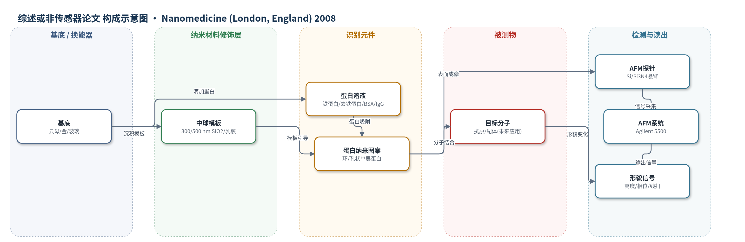
点击图片查看大图 · 依据论文自动绘制
传感器类型
综述或非传感器论文
检测对象
无实际分析物;表面图案化对象:铁蛋白(ferritin)、去铁蛋白(apoferritin)、牛血清白蛋白(BSA)、免疫球蛋白G(IgG)(水溶液/固体表面)
检测原理
该文并非完整传感检测,而是蛋白质表面图案化方法。将蛋白水溶液与单分散二氧化硅或乳胶中球混合后滴于云母、金或玻璃表面,干燥过程中中球自发形成六角密堆模板,蛋白随水弯月面迁移并吸附在球体底部;去离子水冲洗去除中球后,留下环状或孔状蛋白纳米图案。AFM 通过探针与表面接触/轻敲获得形貌、高度和相位信息,用于表征图案周期、直径、厚度和覆盖率。若用于传感,图案化蛋白可作为识别层,目标分子结合后引起局部高度或形貌变化,AFM 可无标记监测,但本文未建立定量检测曲线。
检测灵敏度
原文未报告 LOD、线性范围、灵敏度斜率或相关系数。
效应效果
方法在云母、金和玻璃上均获得周期性蛋白图案。铁蛋白环在云母覆盖率约17%,高12±1 nm,间距550±30 nm;金上覆盖率约12%,环径200±5 nm;玻璃上覆盖率约19%,间距600±30 nm。去铁蛋白玻璃覆盖率约15%,高12±1 nm;IgG云母覆盖率约10%,10×10 µm²内130环,外推1 cm²约1.3亿环,高5±1 nm。BSA高比例形成83%覆盖多孔膜,6×6 µm²内316孔,外推1 cm²约8.77亿孔,厚3.8±0.3 nm;低比例形成26%覆盖环,周期283±16 nm。两周后重复环宽333±17 nm与319±21 nm,误差<6%。未报告选择性、抗干扰、回收率或与ELISA/HPLC/qPCR对比。
传感器的构成
- 基底:云母、模板剥离金薄膜(150 nm Au)或玻璃,提供平面支撑与可选导电/光学表面
- 模板层:单分散二氧化硅或乳胶中球(300/500 nm),干燥时自组装为六角密堆结构并作为掩模
- 功能蛋白层:铁蛋白(ferritin)、去铁蛋白(apoferritin)、BSA、IgG,吸附于模板球底部形成环/孔状纳米图案
- 清洗/去模板步骤:去离子水冲洗,移除中球并暴露周期性蛋白图案
- 读出层:AFM 探针(硅或氮化硅悬臂)与 AFM 系统,用于形貌、高度和相位成像
中文摘要
本文测试了利用单分散中球进行粒子光刻以控制蛋白质表面覆盖率和纳米图案尺寸的方法。单分散二氧化硅或乳胶球在平面基底上干燥时自发形成有序密堆晶层,作为结构模板引导蛋白质在球体底部沉积;随后用去离子水冲洗去除模板球,即可在云母、玻璃和模板剥离金表面获得周期性蛋白质纳米图案。作者以铁蛋白、去铁蛋白、牛血清白蛋白和免疫球蛋白G为模型蛋白,通过改变中球直径和蛋白浓度调控图案形貌与直径。AFM 表征表明蛋白质牢固附着并形成单层厚度结构,环状图案高度与已知蛋白尺寸一致。根据蛋白与中球比例,可得到多孔膜或环状结构。该方法操作简便、通量高、重现性好,可用于表面结合生物测定和生物传感器表面的蛋白质结合相互作用基础研究。
英文摘要
AIMS: The applicability of particle lithography with monodisperse mesospheres is tested with various proteins to control the surface coverage and dimensions of protein nanopatterns.
METHODS & MATERIALS: The natural self-assembly of monodisperse spheres provides an efficient, high-throughput route to prepare protein nanopatterns. Mesospheres assemble spontaneously into organized crystalline layers when dried on flat substrates, which supply a structural frame or template to direct the placement of proteins. The template particles are displaced with a simple rinsing step to disclose periodic arrays of protein nanopatterns on surfaces.
RESULTS & DISCUSSION: The proteins are attached securely to the surface, forming nanopatterns with a measured thickness of a single layer. The morphology and diameter of the protein nanostructures can be tailored by selecting the diameter of the mesospheres and choosing the protein concentration.
CONCLUSIONS: Particle lithography is shown to be a practical, highly reproducible method for patterning proteins on surfaces of mica, glass and gold. High-throughput patterning was achieved with ferritin, apoferritin, bovine serum albumin and immunoglobulin-G. Depending on the ratio of proteins to mesospheres, either porous films or ring structures were produced. This approach can be applied for fundamental investigations of protein-binding interactions of biological systems in surface-bound bioassays and biosensor surfaces.