组成图示
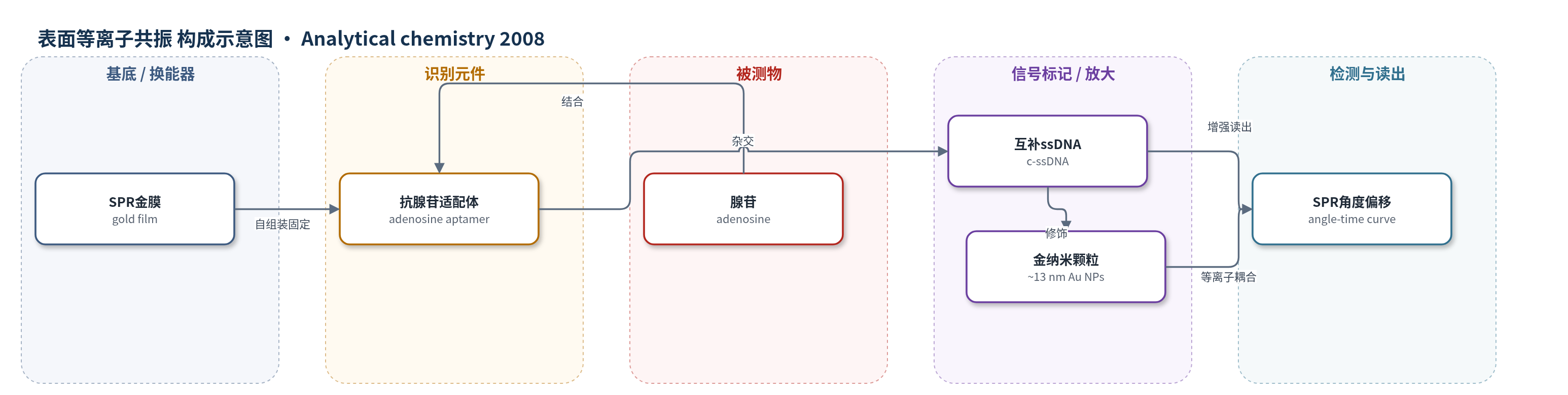
点击图片查看大图 · 依据论文自动绘制
传感器类型
表面等离子共振(SPR)生物传感器
检测对象
腺苷(adenosine),样品基质为50 mM Tris-HCl缓冲液(pH 8.0,含138 mM NaCl)
检测原理
该传感器以抗腺苷适配体为识别元件,通过Au-S键固定在SPR金膜上,并用6-巯基己醇封闭裸露金面。适配体初始呈单链DNA(ss-DNA)结构,可与互补ss-DNA杂交。加入腺苷后,适配体与腺苷特异性结合,由自由卷曲ss-DNA折叠为稳定三级结构;形成三级结构的适配体不能与互补ss-DNA杂交。随后加入金纳米颗粒(Au NPs)标记的互补ss-DNA,只有未结合腺苷的适配体可发生杂交。Au NPs的局域表面等离子体与SPR金膜的表面等离子波发生电子耦合,显著放大SPR角度偏移。腺苷浓度越高,形成三级结构的适配体越多,杂交结合的金纳米颗粒标记互补ss-DNA越少,SPR角度偏移越小。通过记录SPR角度-时间曲线,可获得角度偏移与腺苷浓度对数之间的线性关系。
检测灵敏度
线性范围: 1 × 10⁻⁹–1 × 10⁻⁶ M
效应效果
该SPR传感器对腺苷灵敏度高、选择性好。无腺苷时,Au NPs标记互补ss-DNA杂交产生最大SPR角度偏移约0.831°;1 nM腺苷使偏移降至约0.748°,检测范围为1×10^-9至1×10^-6 M。1 mM尿苷、胞苷、鸟苷仅使偏移轻微下降,1 mM腺苷则显著下降,可区分相似核苷。直接腺苷结合偏移仅约0.004°,需Au NPs放大;1 mM腺苷下仍残留约0.1°杂交信号,归因于适配体构象位阻。作者认为其性能与光学/电化学适配体传感器相当,优于分子印迹SPR方法,可推广至多种小分子。
传感器的构成
- 基底/换能器:SPR金膜(SPR gold film),提供表面等离子共振换能界面
- 识别元件:抗腺苷适配体(anti-adenosine aptamer,5′-SH-C6-AGA GAA CCT GGG GGA GTA TTG CGG AGG AAG GT-3′),经Au-S键固定,结合腺苷后由ss-DNA折叠为三级结构
- 封闭剂:6-巯基己醇(6-mercaptohexan-1-ol),封闭裸露金表面,降低非特异吸附
- 信号标记/放大元件:柠檬酸稳定金纳米颗粒标记互补ss-DNA(Au NPs-tagged complementary ss-DNA,~13 nm Au NPs + 5′-SH-C6-ACC TTC CTC CGC-3′),与未折叠适配体杂交并放大SPR角度偏移
- 缓冲介质:50 mM Tris-HCl(pH 8.0,含138 mM NaCl),维持适配体构象与杂交反应
- 读出系统:Eco Chemie Autolab SPR仪(670 nm激光),记录SPR角度-时间曲线和角度偏移
中文摘要
小分子难以用常规表面等离子共振(SPR)技术直接检测,因为其结合过程引起的折射率变化通常很小。为扩展SPR生物传感器在小分子检测中的应用,作者将适配体技术与金纳米颗粒(Au NPs)的放大效应相结合,设计了一种高灵敏度的SPR传感器。该传感器基于表面抑制检测原理:适配体先以单链DNA(ss-DNA)形式固定在SPR金膜上,此时可与Au NPs标记的互补ss-DNA杂交,引起较大的SPR信号变化;加入腺苷后,适配体由ss-DNA结构折叠为三级结构,无法再与Au NPs标记互补ss-DNA杂交。因此,杂交反应引起的SPR信号变化随具有三级结构适配体数量的增加而降低,而该数量与腺苷浓度成正比。作者以抗腺苷适配体/腺苷体系验证传感性能,结果表明该SPR传感器对腺苷具有良好的灵敏度和高选择性,检测范围为1×10^-9至1×10^-6 M。该策略易于通过更换不同适配体推广到多种小分子的SPR检测,有望为高性能SPR生物传感器设计提供新方向。
英文摘要
Small molecules are difficult to detect by conventional SPR technique directly because the changes in the refractive index resulting from the binding processes of small biomolecules are often small. In order to extend the application of SPR biosensor in detecting a small molecule, we combine the advantage of aptamer technique with the amplifying effect of Au nanoparticles to design a sensitive SPR sensor for detecting small molecules. The principle of this sensor is based on surface inhibition detection. The aptamer is first immobilized on SPR gold film with its ss-DNA structure. The aptamer possessing this structure can be hybridized with Au nanoparticles-tagged complementary ss-DNA and result in a large change of SPR signal. However, the aptamer will change its structure from ss-DNA to tertiary structure after adenosine is added to the SPR cell. The aptamer possessing tertiary structure could not hybridize with Au nanoparticles-tagged complementary ss-DNA. Thus, the change of SPR signal resulted in the hybridization reaction between aptamer and Au nanoparticles-tagged complementary ss-DNA will decrease with the increase of the number of aptamers possessing tertiary structure, which is proportional to the concentration of the small molecule. Based on this principle, we choose a simple system (antiadenosine aptamer/adenosine) to detect the sensing ability of this SPR biosensor for a small molecule. The experimental results confirm that the SPR sensor we developed possesses a good sensitivity and a high selectivity for adenosine. The detection range for adenosine is from 1 x 10 (-9) to 1 x 10 (-6) M. More significantly, it is fairly easy to generalize this strategy to detect a spectrum of small molecules by SPR spectroscopy using different aptamers. Therefore, it is expected that this method may offer a new direction in designing high-performance SPR biosensors for sensitive and selective detection of a wide spectrum of small molecules.