组成图示
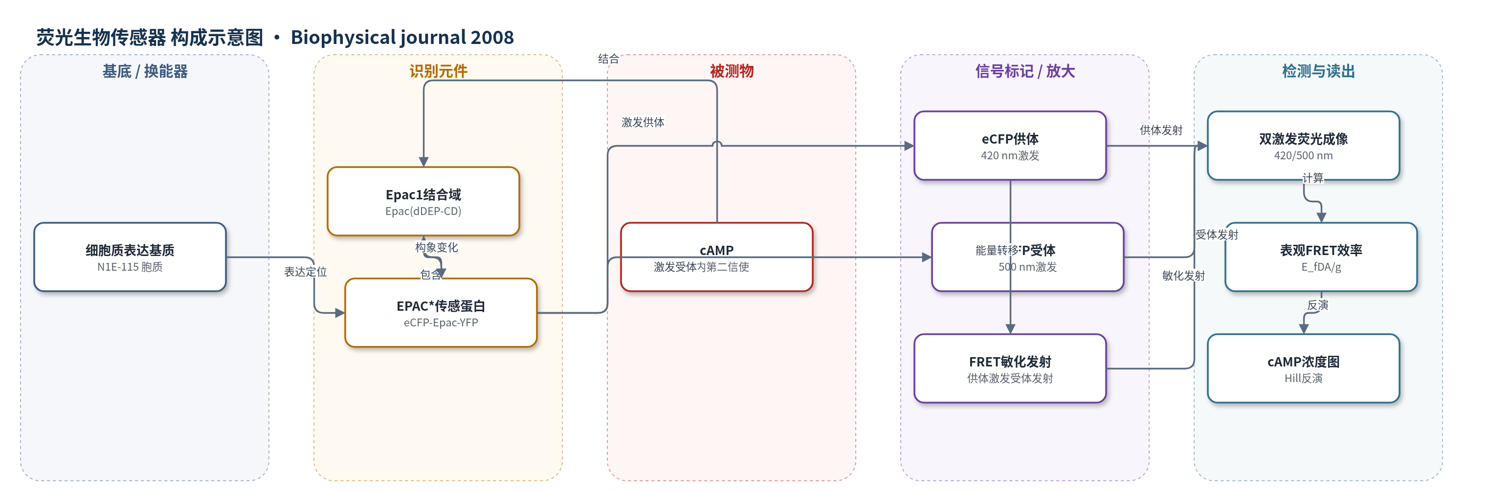
点击图片查看大图 · 依据论文自动绘制
传感器类型
荧光生物传感器
检测对象
环腺苷酸(cAMP, cyclic adenosine monophosphate);样品基质:N1E-115 神经母细胞瘤细胞裂解液与活细胞胞质
检测原理
cAMP 与 EPAC* 中 Epac1 的 cAMP 结合域 Epac(dDEP-CD) 结合后,引起 eCFP 与 eYFP 之间距离和取向变化,从而改变 FRET 效率。低 cAMP 时 FRET 效率约 35%±2%,高 cAMP 时降至 20%±2%,ΔE 约 15%±1%。检测采用双激发波长:420 nm 激发 eCFP,同时检测 eCFP 发射和 eYFP 敏化发射;500 nm 激发 eYFP,检测 eYFP 发射。通过表观 FRET 效率 E_fDA/g 校正受体直接激发和供体串扰,避免 eYFP 受 pH/离子环境引起的非 FRET 强度变化干扰。校准曲线用 Hill 方程拟合,EC50 约 1.5±0.2 μM,nH 约 -0.95±0.05,再反演得到 cAMP 浓度。该方法无酶放大,信号直接来自 FRET 效率变化。
检测灵敏度
定量范围: 150 nM–15 μM;EC50: (1.5 ± 0.2) μM;nH: -0.95 ± 0.05;ΔE: 15% ± 1%
效应效果
与传统 eYFP/eCFP 强度比相比,E_fDA/g 对 pH 6.5–8 的离子环境变化不敏感,不同细胞批次校准曲线的 pmax/p0 变化小于 1%,而强度比变化大于 7%,抗干扰和批次稳定性更好。校准得到 EC50=1.5±0.2 μM、nH=-0.95±0.05,FRET 效率变化 ΔE=15%±1%。在共表达 5-HT7 受体的 N1E-115 细胞中,1 mM 5-CT 刺激使胞内 cAMP 从基础 0.32±0.62 μM 升至 1.62±1.83 μM;活性微域可升至 9 μM,周围被动区域低于 2 μM。5-HT7 特异性拮抗剂 SB269970 可完全阻断激动剂诱导的 cAMP 升高,显示受体特异性。作者认为该方法适用于活细胞高时空分辨 cAMP 定量及其他 1:1 FRET 传感器。
传感器的构成
- 识别元件:Epac1 cAMP 结合结构域 Epac(dDEP-CD),结合 cAMP 并诱导构象变化
- 供体荧光标记:增强青色荧光蛋白 eCFP,融合于 Epac1 N 端,作为 FRET 供体
- 受体荧光标记:增强黄色荧光蛋白 eYFP,融合于 Epac1 C 端,作为 FRET 受体
- 传感融合蛋白:EPAC*(eCFP-Epac(dDEP-CD)-eYFP),1:1 供体/受体化学计量,胞质定位
- 表达基质:N1E-115 神经母细胞瘤细胞胞质,作为 EPAC* 表达与 cAMP 检测环境
- 信号读出:双激发波长荧光光谱/显微镜(420 nm 与 500 nm),检测 eCFP/eYFP 发射与敏化发射
中文摘要
基于 Förster 共振能量转移(FRET)的生物传感器可用于定量分析细胞内信号,包括环腺苷酸(cAMP)监测,受到广泛关注。串联生物传感器在供体激发波长下测量供体/受体发射比是常用方法,但该比值不仅随 cAMP 浓度变化,还会受离子环境或其他影响荧光蛋白折叠概率的因素影响。本文采用基于两种激发波长的光谱 FRET 分析方法,利用“cAMP 直接激活的交换蛋白”(Epac)实现高时间分辨率和空间分辨率的绝对 cAMP 浓度可靠测量。该方法简化了 FRET 分析,无需额外校准程序。由 cAMP 浓度变化引起的 FRET 效率(E)变化为 ΔE=15%,E 在低 cAMP 时为 35%,高 cAMP 时为 20%,从而可在 150 nM 至 15 μM 范围内定量测量 cAMP 浓度。该方法也适用于具有 1:1 供体/受体化学计量的其他 FRET 生物传感器。作为原理验证,作者在活细胞内测量了空间分辨的 cAMP 浓度,并确定了 Gs 偶联 5-羟色胺受体亚型 7(5-HT7)刺激后 cAMP 水平的动态变化。
英文摘要
Förster resonance energy transfer (FRET)-based biosensors for the quantitative analysis of intracellular signaling, including sensors for monitoring cyclic adenosine monophosphate (cAMP), are of increasing interest. The measurement of the donor/acceptor emission ratio in tandem biosensors excited at the donor excitation wavelength is a commonly used technique. A general problem, however, is that this ratio varies not only with the changes in cAMP concentration but also with the changes of the ionic environment or other factors affecting the folding probability of the fluorophores. Here, we use a spectral FRET analysis on the basis of two excitation wavelengths to obtain a reliable measure of the absolute cAMP concentrations with high temporal and spatial resolution by using an "exchange protein directly activated by cAMP". In this approach, FRET analysis is simplified and does not require additional calibration routines. The change in FRET efficiency (E) of the biosensor caused by [cAMP] changes was determined as DeltaE = 15%, whereas E varies between 35% at low and 20% at high [cAMP], allowing quantitative measurement of cAMP concentration in the range from 150 nM to 15 microM. The method described is also suitable for other FRET-based biosensors with a 1:1 donor/acceptor stoichiometry. As a proof of principle, we measured the specially resolved cAMP concentration within living cells and determined the dynamic changes of cAMP levels after stimulation of the Gs-coupled serotonin receptor subtype 7 (5-HT7).