组成图示
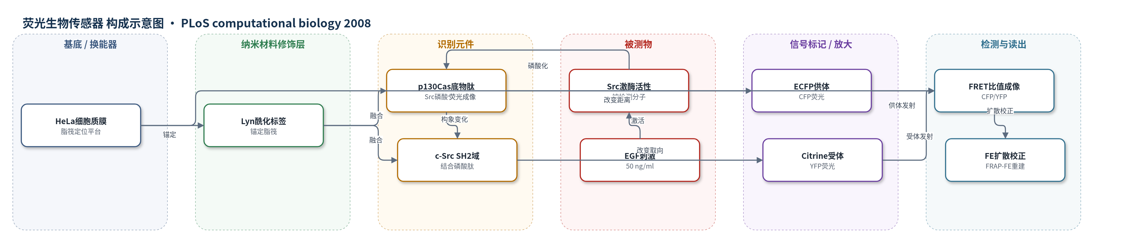
点击图片查看大图 · 依据论文自动绘制
传感器类型
荧光生物传感器
检测对象
Src 激酶活性(Src kinase activity);样品基质:HeLa 活细胞质膜脂筏(EGF 刺激)
检测原理
该传感器为遗传编码 FRET 生物传感器:Src 激酶磷酸化其内部 p130Cas 来源底物肽,磷酸肽与同源 c-Src SH2 结构域结合,使 ECFP 与 Citrine 的距离或相对取向改变,从而改变 FRET 效率;通常以 CFP/YFP 发射比升高表示 Src 活性增强。由于生物传感器在质膜脂筏或胞质中可扩散,表观 FRET 图像同时包含原位激酶激活和已激活传感器迁移带来的信号。作者先用 FRAP 估计表观扩散系数,再用有限元模型模拟相邻时间步的扩散贡献,并从 FRET 比值图像中扣除,得到扩散校正后的 Src 激活图。脂筏锚定的 Lyn-Src 主要受二维扩散支配,EGF 刺激后高 Src 活性集中于细胞周边脂筏簇且位置相对静止。
检测灵敏度
未报告LOD、线性范围、灵敏度斜率或检测相关系数。
效应效果
FRAP 结果显示,Lyn-Src 在脂筏的表观扩散系数为 0.11±0.01 μm²/s(n=43),KRas-Src 为 0.18±0.02 μm²/s(n=17),胞质型 Src 为 0.93±0.06 μm²/s;MbCD 破坏脂筏后 Lyn-Src 升至 0.17±0.01 μm²/s,KRas-Src 为 0.20±0.01 μm²/s。扩散模型决定系数 R² 分别为 Lyn-Src 0.79±0.033(n=50)、KRas-Src 0.56±0.06(n=15),MbCD 处理后为 0.68±0.07 和 0.47±0.06;胞质型 R²=0.33±0.1(n=5)。扩散校正后的 FRET 图像显示 EGF 刺激下脂筏细胞周边出现离散高 Src 活性簇且位置相对静止,与固定细胞免疫染色一致。作者认为该方法可推广重建其他信号分子的活细胞时空激活图。
传感器的构成
- 荧光供体层:单体 ECFP(文中简称 CFP),作为 FRET 供体,发射被 Citrine 接受。
- 荧光受体层:Citrine(改进型 EYFP,文中简称 YFP),作为 FRET 受体,与 ECFP 构成 FRET 对。
- 识别响应层:p130Cas 来源 Src 底物肽,被 Src 磷酸化后触发构象变化。
- 识别结合层:c-Src SH2 结构域,结合磷酸化底物肽并改变 ECFP/Citrine 距离或取向。
- 膜定位修饰层:Lyn 激酶 N 端肉豆蔻酰化/棕榈酰化序列,将 Lyn-Src 生物传感器锚定于质膜脂筏。
- 膜定位修饰层:KRas C 端法尼基化序列,将 KRas-Src 生物传感器定位于非脂筏质膜区室。
- 表达与换能平台:HeLa 活细胞质膜/脂筏,作为生物传感器定位与 Src 活性发生的微区室。
- 读出与校正模块:荧光显微镜 FRET 比值成像结合 FRAP-FE 扩散校正,输出 Src 激活时空图。
中文摘要
基于荧光共振能量转移(FRET)的遗传编码生物传感器已被广泛用于以高时空分辨率可视化活细胞中的分子活性,但生物传感器蛋白的快速扩散会阻碍真实分子激活图的精确重建。作者基于荧光漂白后恢复(FRAP)实验,发展了一种有限元(FE)方法,用于分析、模拟并扣除可移动生物传感器的扩散效应。该方法被应用于分析定位于不同亚细胞区室的 Src FRET 生物传感器在活细胞中的迁移性。结果显示,胞质型 Src 生物传感器的迁移速度比锚定于质膜不同区室的生物传感器快 4–8 倍(0.93±0.06 μm²/s;脂筏 0.11±0.01 μm²/s,脂筏外 0.18±0.02 μm²/s)。脂筏上的生物传感器迁移慢于脂筏外,且主要受二维扩散支配。将该扩散效应从 FRET 比值图像中扣除后,在靠近细胞周边的脂筏簇区观察到较高的 Src 活性,且经表皮生长因子(EGF)刺激后位置相对静止。结果表明 EGF 诱导脂筏处 Src 激活具有协调的时空模式。该 FE 方法也为研究活细胞中信号分子迁移和重建时空激活图提供了集成图像分析平台。
英文摘要
Genetically encoded biosensors based on fluorescence resonance energy transfer (FRET) have been widely applied to visualize the molecular activity in live cells with high spatiotemporal resolution. However, the rapid diffusion of biosensor proteins hinders a precise reconstruction of the actual molecular activation map. Based on fluorescence recovery after photobleaching (FRAP) experiments, we have developed a finite element (FE) method to analyze, simulate, and subtract the diffusion effect of mobile biosensors. This method has been applied to analyze the mobility of Src FRET biosensors engineered to reside at different subcompartments in live cells. The results indicate that the Src biosensor located in the cytoplasm moves 4-8 folds faster (0.93+/-0.06 microm(2)/sec) than those anchored on different compartments in plasma membrane (at lipid raft: 0.11+/-0.01 microm(2)/sec and outside: 0.18+/-0.02 microm(2)/sec). The mobility of biosensor at lipid rafts is slower than that outside of lipid rafts and is dominated by two-dimensional diffusion. When this diffusion effect was subtracted from the FRET ratio images, high Src activity at lipid rafts was observed at clustered regions proximal to the cell periphery, which remained relatively stationary upon epidermal growth factor (EGF) stimulation. This result suggests that EGF induced a Src activation at lipid rafts with well-coordinated spatiotemporal patterns. Our FE-based method also provides an integrated platform of image analysis for studying molecular mobility and reconstructing the spatiotemporal activation maps of signaling molecules in live cells.