组成图示
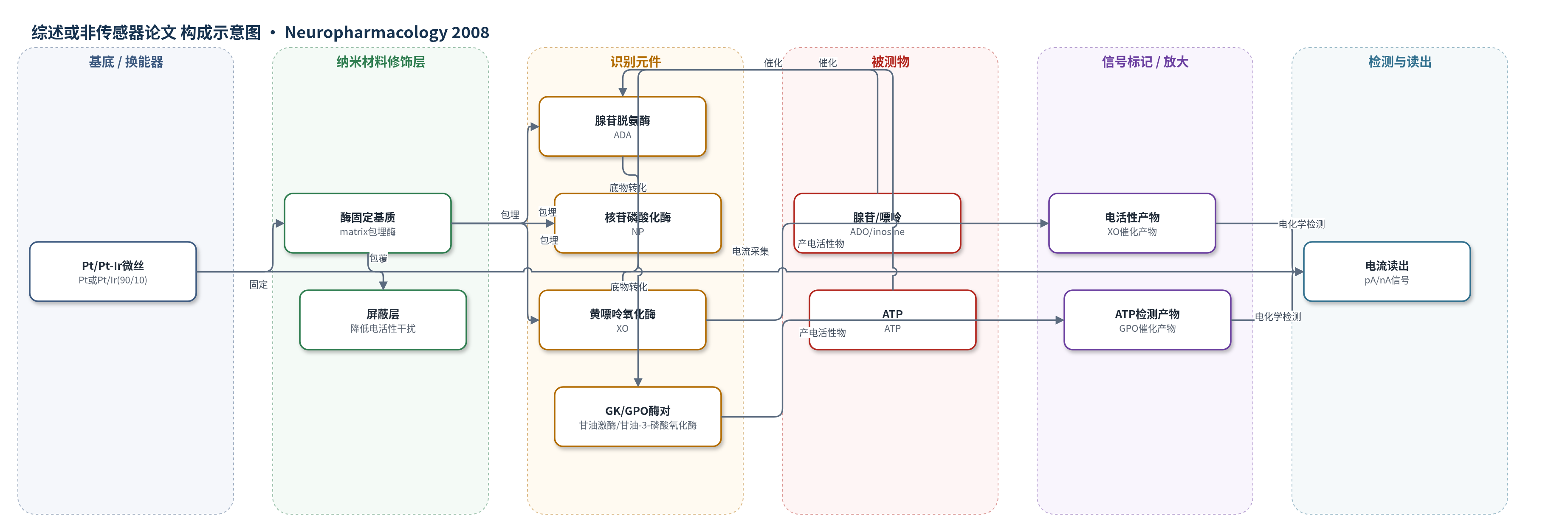
点击图片查看大图 · 依据论文自动绘制
传感器类型
综述或非传感器论文
检测对象
腺苷(adenosine, ADO)、肌苷/次黄嘌呤等嘌呤(purines)、ATP;样品基质:大鼠小脑/海马脑片细胞外灌流液。
检测原理
该研究使用电化学微电极生物传感器检测脑片细胞外嘌呤。腺苷传感器中,腺苷脱氨酶(ADA)将腺苷转化为肌苷,核苷磷酸化酶(NP)将肌苷转化为次黄嘌呤,黄嘌呤氧化酶(XO)氧化次黄嘌呤产生电活性产物,在Pt/Pt-Ir微丝上产生与腺苷/嘌呤浓度成正比的电流;屏蔽层降低多巴胺、5-HT、抗坏血酸等干扰。ATP传感器中,甘油激酶(GK)在甘油共底物存在下将ATP磷酸化为ADP和甘油-3-磷酸,甘油-3-磷酸氧化酶(GPO)氧化甘油-3-磷酸产生电活性信号。POM-1抑制NTPDase后,ATP向ADP/腺苷转化减少,腺苷传感器电流下降,ATP传感器因局部ATP分解减少而电流略升。
检测灵敏度
原文未报告LOD、线性范围、灵敏度斜率或相关系数。
效应效果
POM-1抑制小脑片ATP分解强于ARL 67156:100 μM POM-1对100 μM ATP抑制57.4±3.8%,ARL 67156为13.9±4.3%;对25 μM ATP抑制68±7%,ARL 67156为43±7.5%。HPLC显示其阻断ATP→ADP第一步且可逆。腺苷传感器显示POM-1使ATP产生的腺苷由4.8±0.7 μM降至2.3±0.2 μM(约52%),洗脱恢复;ATP传感器检测ATP由7.2±3%升至8.7±2%。但POM-1脱靶抑制突触传递:小脑平行纤维fEPSP 10 min降低50.8±6.9%,长时间85–95%;攀纤维降低75.5±15%;海马CA1降低88.9±1.4%。配对脉冲比升高、突触前波幅降低提示突触前机制;P2拮抗剂不能阻断,ATP传感器未检出ATP积累,说明非NTPDase抑制所致。
传感器的构成
- 换能器电极:Pt或Pt/Ir(90/10)微丝,蚀刻至25–50 μm,暴露长度500 μm,用于电化学检测。
- 酶固定层:未指明具体聚合物的matrix,沉积于电极周围,包埋酶并允许底物扩散。
- 识别/催化元件(ADO传感器):腺苷脱氨酶(ADA)、核苷磷酸化酶(NP)、黄嘌呤氧化酶(XO),将腺苷/嘌呤逐步转化为电活性产物。
- 屏蔽层:screening layer,降低5-HT、多巴胺、去甲肾上腺素、抗坏血酸等非特异电活性干扰。
- 对照空传感器:含matrix但无酶,用于扣除非特异电活性干扰。
- ATP传感器催化元件:甘油激酶(GK)与甘油-3-磷酸氧化酶(GPO),在甘油存在下将ATP转化为可检测产物。
- 共底物:甘油(2 mM),作为ATP检测所需共底物。
中文摘要
嘌呤能信号研究需要强效且亚型选择性的NTPDase抑制剂,以阻断细胞外ATP/ADP代谢。ARL 67156是目前常用抑制剂,但效力有限。聚氧钨酸钠(POM-1)是新型NTPDase抑制剂,对重组NTPDase 1、2、3均有抑制作用。本研究在小脑和海马脑片中评估POM-1。孔雀绿磷酸盐测定、HPLC和生物传感器显示,POM-1比ARL 67156更能阻断小脑片ATP分解,作用位点为ATP转化为ADP的第一步,且可逆。但POM-1对突触传递有脱靶作用:在小脑平行纤维-浦肯野细胞突触,少数突触先出现约20%的EPSP短暂升高,提示突触前A1受体张力降低;随后出现持续抑制,且阻断腺苷和ATP受体后仍存在,说明并非NTPDase抑制所致。配对脉冲比升高和突触前波幅降低提示其减少谷氨酸释放。POM-1在攀纤维和海马CA1突触也产生类似抑制。因此,POM-1虽优于ARL 67156,但突触抑制限制其应用。
英文摘要
Understanding the mechanisms and properties of purinergic signalling would be greatly assisted by the discovery of subtype selective and potent inhibitors of the NTPDase enzymes, which metabolise nucleotides such as ATP and ADP in the extracellular space. Currently ARL 67156 is the best available NTPDase inhibitor, but its relatively poor efficacy means that negative results are difficult to interpret. POM-1 (sodium polyoxotungstate) is a novel NTPDase inhibitor, which has shown promising results with the inhibition of recombinant NTPDases 1, 2 and 3. We have tested the effectiveness and physiological effects of POM-1 with cerebellar and hippocampal slices. Using the malachite green phosphate assay, HPLC and biosensor measurements we have found that POM-1 is more effective at blocking ATP breakdown in cerebellar slices than ARL 67156. The site of inhibition is at the first step of the breakdown cascade (conversion of ATP to ADP) and the effects of POM-1 appear readily reversible. However, POM-1 has multiple effects on synaptic transmission. At the cerebellar parallel fibre-Purkinje cell (PF) synapse POM-1 produced a long lasting inhibition of transmission, which was preceded in a minority of synapses by a transient increase in PF excitatory postsynaptic potential (EPSP) amplitude (approximately 20%). This increase in PF EPSP amplitude appears to result from a reduction in the tonic activation of presynaptic A1 receptors, consistent with POM-1 preventing the breakdown of ATP to adenosine. The reduction in PF EPSP amplitude does not however appear to result from NTPDase inhibition as it persists when both adenosine and ATP (P2Y and P2X) receptors are blocked. An increase in paired pulse ratio and a reduction in presynaptic volley amplitude suggest that there is a presynaptic component of POM-1 action which reduces glutamate release. POM-1 produced similar inhibition at climbing fibre synapses and at hippocampal CA1 pyramidal synapses. Thus although POM-1 is more effective than ARL 67156 at blocking ATP breakdown its usefulness is limited by off-target actions on synaptic transmission.