组成图示
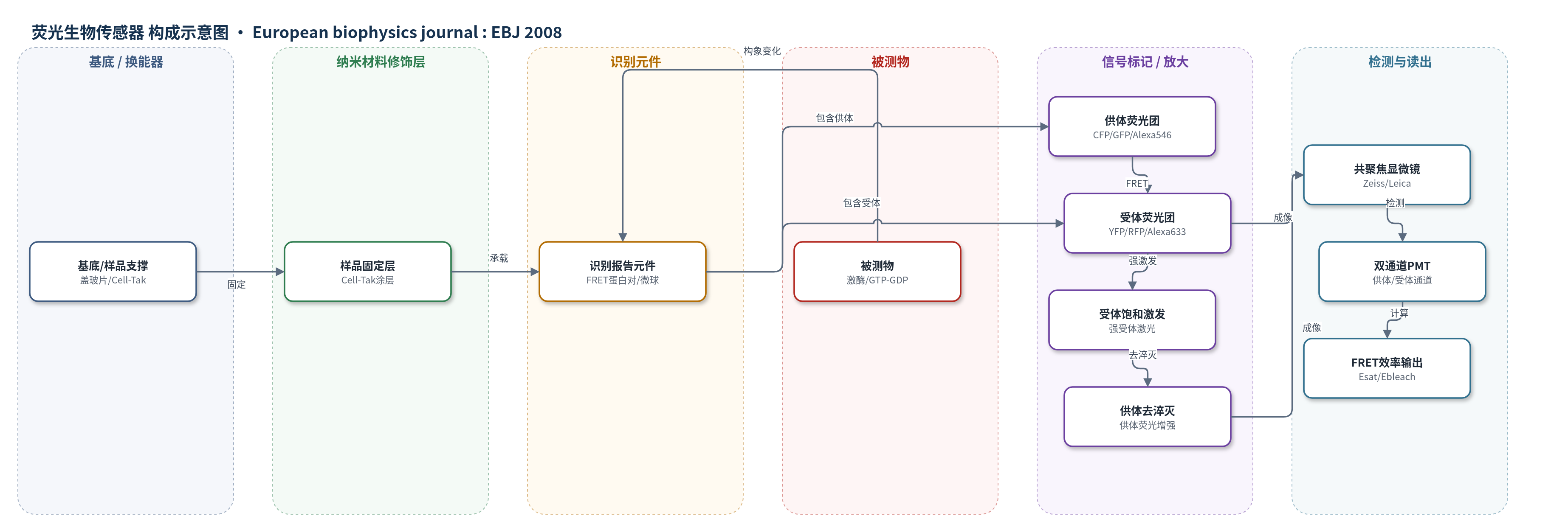
点击图片查看大图 · 依据论文自动绘制
传感器类型
荧光生物传感器
检测对象
FRET 供体-受体对(Alexa 546-Alexa 633,PBS 中的生物素聚苯乙烯微球)、蛋白激酶C磷酸化/活性(protein kinase C, PKC;CKAR CFP-YFP 报告子,固定 CHO 细胞)、GTP/GDP 转换/小 GTPase 活性(GTP/GDP turnover, Raichu Cdc42/Rac1/RhoA,活 Jurkat T 细胞);样品基质为 PBS 微球、固定 CHO 细胞、活 Jurkat T 细胞(RPMI1640/FCS)。
检测原理
供体-受体荧光蛋白对(CFP-YFP、GFP-Raichu-RFP)距离在 2–10 nm 时发生 FRET,效率 E=kt/(k_d^-+kt),随距离六次方变化。被测物结合或磷酸化使报告子构象改变,改变供体-受体距离,从而改变供体淬灭程度。satFRET 用低强度激发供体、高强度激发受体,使受体激发态饱和;饱和受体不再满足 FRET 光谱重叠,供体发生去淬灭,供体荧光增强。比较受体未饱和与饱和时的供体强度,并结合受体饱和度 asat,按 Esat=1-asat/(IDonorSat/IDonor+asat-1) 计算效率;方法可逆,可加光漂白校正,实现时间分辨读出。
检测灵敏度
—
效应效果
在 Alexa 546-Alexa 633 微球中,satFRET 效率随供体/受体化学计量比变化,与受体光漂白法呈清晰相关,与敏化发射法一致性较差。CHO 细胞 CFP-YFP 实验中,最高受体激发下 asat=0.31±0.02,漂白前 Esat=0.32,受体光漂白法 Ebleach=0.35;漂白后仍可重复测量,漂白区效率下降而周围区域仍可读出。活 T 细胞实验中 asat=0.62±0.01,受体信号约损失 40%,经光漂白校正的 Esat 随激活时间上升,并与固定细胞光漂白结果良好对应;完全漂白后 E0sat=-0.02±0.01 作为零点。方法仅需两帧图像,约 0.1 s 出结果,可重复,适合常规共聚焦显微镜。
传感器的构成
- 基底/样品支撑:盖玻片或 LabTek 腔室盖玻片,用于承载微球或细胞样品。
- 样品固定层:Cell-Tak 涂层,通过吸附固定生物素聚苯乙烯微球或细胞。
- 识别/报告元件:基因编码 FRET 荧光蛋白对(CFP-YFP、GFP-Raichu-RFP)或 Alexa 546-streptavidin/Alexa 633-streptavidin 修饰的生物素聚苯乙烯微球,响应构象/化学计量变化。
- 供体荧光团:CFP、GFP 或 Alexa 546,接受激发并向受体转移能量。
- 受体荧光团:YFP、RFP 或 Alexa 633,接受 FRET 能量并在强激发下进入激发态饱和。
- 信号标记/放大:无外源标记,利用受体饱和导致供体去淬灭的可逆荧光变化作为信号。
- 换能/读出:共聚焦激光扫描显微镜(Zeiss 510/510 Meta、Leica SP2 AOBS)与 PMT 双通道检测供体/受体荧光。
中文摘要
本文从理论和实验上证明,可通过直接且系统地饱和受体分子激发态来定量 Förster 共振能量转移(FRET),并将该受体耗竭方法称为 satFRET。该方法可逆,适合时间分辨测量。作者基于供体-受体分子状态微分方程组的稳态解建立模型,并纳入测量过程中受体光漂白的影响。实验采用不同化学计量比的 Alexa 546-Alexa 633 微球,在共聚焦显微镜中验证,将激发态饱和得到的能量转移效率与敏化发射和受体光漂白法比较。结果形成可在常规共聚焦显微镜上对固定和活细胞进行时间分辨 FRET 测量的方案,并应用于含 CFP-YFP 对的固定 CHO 细胞;在活 T 细胞激活实验中,利用编码 GFP-RFP 的 GTP/GDP 转换生物传感器比较 satFRET 与固定细胞受体光漂白结果,展示了时间分辨能力。
英文摘要
We demonstrate theoretically and experimentally the quantification of Förster resonance energy transfer (FRET) by direct and systematic saturation of the excited state of acceptor molecules. This version of acceptor depletion methods for FRET estimation, denoted as "satFRET" is reversible and suitable for time-resolved measurements. The technique was investigated theoretically using the steady-state solution of the differential equation system of donor and acceptor molecular states. The influence of acceptor photobleaching during measurement was included in the model. Experimental verification was achieved with the FRET-pair Alexa 546- Alexa 633 loaded on particles in different stoichiometries and measured in a confocal microscope. Estimates of energy transfer efficiency by excited state saturation were compared to those obtained by measurements of sensitised emission and acceptor photobleaching. The results lead to a protocol that allows time-resolved FRET measurements of fixed and living cells on a conventional confocal microscope. This procedure was applied to fixed Chinese hamster ovary cells containing a cyan fluorescent protein and yellow fluorescent protein pair. The time resolution of the technique was demonstrated in a live T cell activation assay comparing the FRET efficiencies measured using a genetically encoded green and red fluorescent protein biosensor for GTP/GDP turnover to those measured by acceptor photobleaching of fixed cells.