组成图示
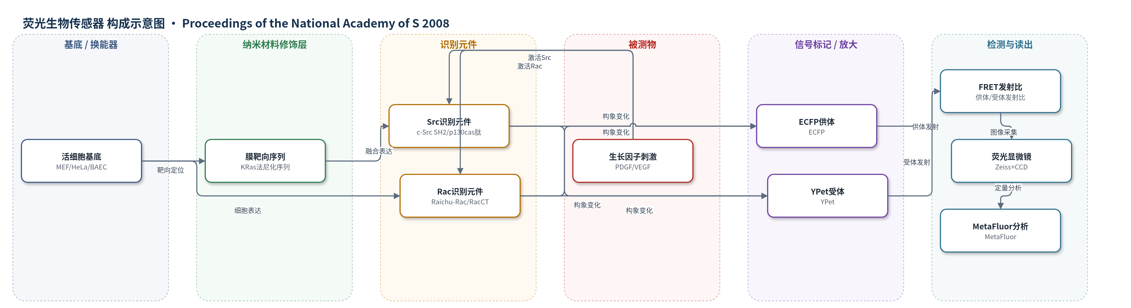
点击图片查看大图 · 依据论文自动绘制
传感器类型
荧光生物传感器
检测对象
Src 激酶活性(Src kinase activity)、Rac GTPase 活性(Rac GTPase activity)、钙离子(Ca2+)、MT1-MMP 蛋白酶活性(MT1-MMP activity);样品基质:活细胞(HeLa、MEF、BAEC)及体外纯化蛋白溶液。
检测原理
FRET 生物传感器由 ECFP 供体、YPet 受体和识别元件串联组成。当 Src 磷酸化 p130cas 来源底物肽时,c-Src SH2 域结合磷酸肽,引起传感器构象变化,改变 ECFP 与 YPet 的距离或取向;Rac 结合 GTP 时改变 Raichu-Rac 构象;Ca2+ 结合钙调蛋白/钙结合元件;MT1-MMP 切割底物肽。由于 FRET 效率与供体-受体距离的六次方成反比,构象变化导致供体发射降低、受体发射升高,供体/受体发射比发生可定量变化。YPet 与 ECFP 具有较高 FRET 效率及弱二聚倾向,提高动态范围,使信号随被测物活性或浓度增加而增强。
检测灵敏度
原文未报告 LOD、线性范围、灵敏度斜率或相关系数;报告 FRET 动态范围变化:Src 体外 120%(ECFP/YPet)vs 25%(ECFP/Citrine);Rac 46% vs 28%/26%;MT1-MMP 570% vs 100%/90%;Ca2+ 350%/65%/135%;活细胞 Src 176%/77%/32%;Rac 70%/15%/5%;Ca2+ 253% vs 242%。
效应效果
在活细胞中,ECFP/YPet 显著提高传感器动态范围:Src 为 176%,高于 cpVenus 77% 和 Citrine 32%;Rac 为 70%,高于 Venus 15% 和 cpVenus 5%;Ca2+ 为 253%,与 cpVenus 242% 相当且绝对发射比更大;MT1-MMP 仅 YPet 版本对 10% FBS 刺激显示最大响应。VEGF 诱导的 Src 激活可被 VEGFR2 抑制剂 SU1498 和 Src 抑制剂 PP1 消除,说明响应具有信号特异性。PDGF 诱导的 Rac 极化可被 PP1 或 Src/Yes/Fyn 三重敲除阻断;Src 全局激活可被 RacN17 或 Cyto D 抑制,并被 RacV12 增强,显示 Src 与 Rac 相互调节。作者主张 ECFP/YPet 可作为通用 FRET 对开发高灵敏活细胞传感器。
传感器的构成
- 细胞基底层:活细胞(MEF/HeLa/BAEC),作为传感器表达与信号发生环境。
- 识别元件层:c-Src SH2 域与 p130cas 来源底物肽,识别 Src 磷酸化事件并触发构象变化。
- 识别元件层:Raichu-Rac/RacCT 元件,识别 Rac GTPase 活性状态。
- 识别元件层:YC3.6 钙结合元件与 proMMP-2 来源底物肽 CPKESCNLFVLKD,分别识别 Ca2+ 和 MT1-MMP 切割。
- 供体荧光蛋白层:ECFP,作为 FRET 供体,其发射随与受体距离/取向变化。
- 受体荧光蛋白层:YPet(对照 Citrine/cpVenus/Venus),作为 FRET 受体,提高 FRET 效率。
- 膜靶向层:KRas 法尼化底物序列 KKKKKKSKTKCVIM,将 Src 传感器靶向质膜。
- 表达/展示层:pRSETB、pcDNA3.1 或 pDisplay 载体,用于细菌/哺乳动物细胞表达或外膜展示。
- 信号读出层:荧光显微镜与 MetaFluor 软件,采集 ECFP/YPet 发射并计算 FRET 发射比。
中文摘要
基于荧光共振能量转移(FRET)的基因编码生物传感器能够以高时空分辨率在活细胞中可视化信号事件,但其灵敏度有限,限制了广泛应用。本研究将增强型青色荧光蛋白(ECFP)与黄色荧光蛋白变体 YPet 配对,构建 ECFP/YPet FRET 对。该 FRET 对可显著提高多种信号分子生物传感器的灵敏度,包括酪氨酸激酶 Src、小 GTPase Rac、钙离子和膜结合基质金属蛋白酶 MT1-MMP,且无需对每个传感器进行专门优化。利用改进后的传感器,作者观察到 PDGF 诱导的 Src 与 Rac 激活在微图案表面定向迁移细胞中呈现不同亚细胞模式:Rac 活性高度极化并富集于细胞前缘,而 Src 活性相对均匀。这些 FRET 传感器还揭示 Src 与 Rac 相互调节。结果表明,同一信号反馈环路中的分子可在不同亚细胞位置受到差异调节。ECFP/YPet 可作为通用 FRET 对,用于开发高灵敏生物传感器,以解析活细胞亚细胞位置的分子层级。
英文摘要
Genetically encoded biosensors based on FRET have enabled the visualization of signaling events in live cells with high spatiotemporal resolution. However, the limited sensitivity of these biosensors has hindered their broad application in biological studies. We have paired enhanced CFP (ECFP) with YPet, a variant of YFP. This ECFP/YPet FRET pair markedly enhanced the sensitivity of biosensors (several folds enhancement without the need of tailored optimization for each individual biosensor) for a variety of signaling molecules, including tyrosine kinase Src, small GTPase Rac, calcium, and a membrane-bound matrix metalloproteinase MT1-MMP. The application of these improved biosensors revealed that the activations of Src and Rac by PDGF displayed distinct subcellular patterns during directional cell migration on micropatterned surface. The activity of Rac is highly polarized and concentrated at the leading edge, whereas Src activity is relatively uniform. These FRET biosensors also led to the discovery that Src and Rac mutually regulate each other. Our findings indicate that molecules within the same signaling feedback loop can be differentially regulated at different subcellular locations. In summary, ECFP/YPet may serve as a general FRET pair for the development of highly sensitive biosensors to allow the determination of molecular hierarchies at subcellular locations in live cells.