组成图示
点击图片查看大图 · 依据论文自动绘制
传感器类型
荧光生物传感器
检测对象
游离 Gαγ(Gαγ)、PI(3,4,5)P3(磷脂酰肌醇-3,4,5-三磷酸);样品基质为活细胞(HEK293、RBL-2H3)质膜附近胞质,刺激物为 fMLP 或 IB-MECA。
检测原理
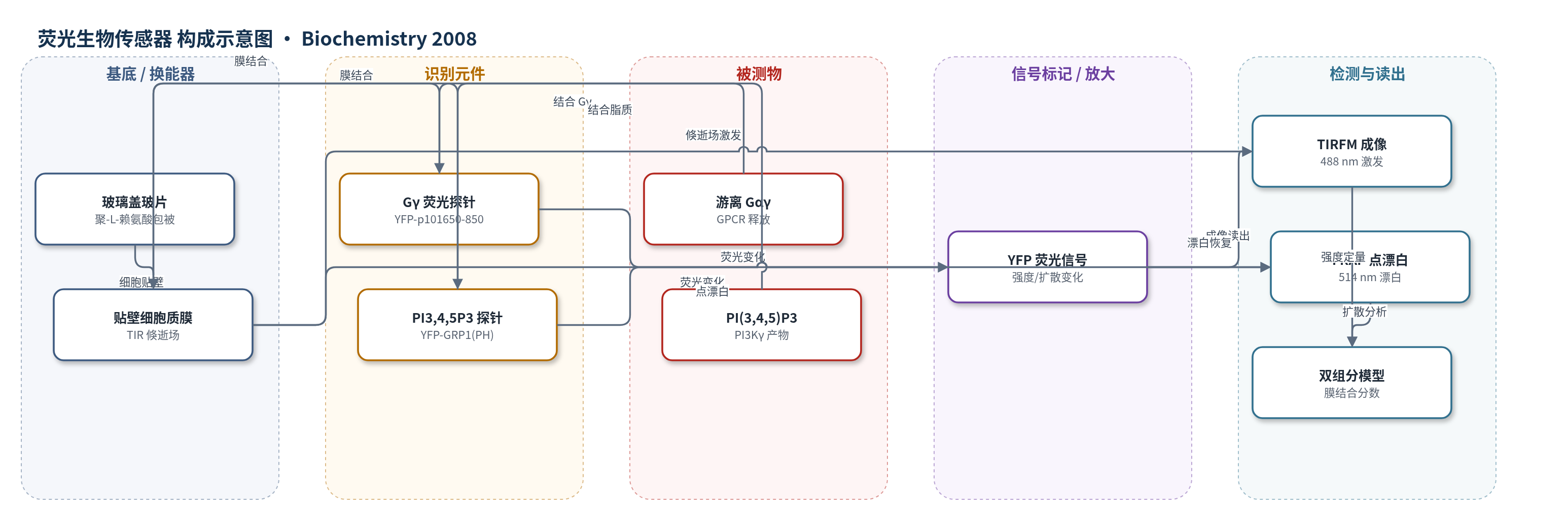
GPCR 被 fMLP 或 IB-MECA 激活后释放游离 Gαγ。YFP-p101650-850 的 p101 Gαγ 结合域与 Gαγ 结合,由胞质转位至质膜;PI3Kγ 被 Gαγ 招募并催化生成 PI(3,4,5)P3,YFP-GRP1(PH) 的 PH 域结合该脂质并转位至质膜。TIR 倏逝场仅激发质膜附近约 100–150 nm 的荧光分子,因此膜结合增加表现为 TIR 荧光强度上升。FRAP 点漂白后,胞质快扩散组分与膜结合慢扩散组分共同恢复;膜结合分数增加、平均扩散系数下降。通过全局双组分模型提取膜结合/胞质振幅,并结合倏逝场强度分布计算膜富集倍数,比较两个生物传感器即可估算 Gαγ 释放到 PI(3,4,5)P3 形成的信号放大。
检测灵敏度
未报告 LOD、线性范围、灵敏度斜率或相关系数。
效应效果
与 CLSM 相比,TIR/FRAP 能检测受体刺激后 YFP-p101650-850 的弱质膜转位。HEK293 细胞中,未刺激时约 17% 的 YFP-p101650-850 位于 TIR 体积内质膜,1 µM fMLP 刺激后升至约 31%;YFP-GRP1(PH) 由约 60% 升至约 90%。膜富集倍数分别为 3.5±0.5 至 7.5±0.7 和 35.5±1.7 至 189±29,估算信号放大约 25 倍。RBL-2H3 细胞中放大约 5–10 倍,表达 p87PIKAP 的 HEK293 细胞约 2.5 倍。两种生物传感器对 fMLP 的 EC50 约 0.2 nM。作者认为该方法可推广至多种转位荧光生物传感器和活细胞信号转导定量。
传感器的构成
- 细胞培养基底:聚-L-赖氨酸(poly-L-lysine)包被玻璃盖玻片,用于 HEK293 或 RBL-2H3 细胞贴壁。
- 换能/成像基底:玻璃盖玻片与高数值物镜形成 TIR 倏逝场,选择性激发质膜附近约 100–150 nm 的荧光分子。
- 识别元件:YFP-p101650-850(p101 的 Gαγ 结合域)识别游离 Gαγ;YFP-GRP1(PH)(GRP1 PH 域)识别 PI(3,4,5)P3。
- 信号标记物:YFP 荧光蛋白,结合后发生胞质-质膜转位,改变 TIR 荧光强度与扩散系数。
- 读出装置:TIRFM 成像(488 nm 激发)与 514 nm 点漂白 FRAP,EM-CCD/CCD 记录,卷积双组分模型定量膜结合分数。
中文摘要
磷脂酰肌醇 3-激酶 γ(PI3Kγ)在 G 蛋白偶联受体(GPCR)刺激后通过 Gαγ 释放而被激活,其调节亚基 p101 或 p87 与 Gαγ 相互作用,将酶招募至质膜。然而,受体介导的 Gαγ 释放不足以使 p101 发生可用经典荧光显微镜观察的转位。由于质膜结合蛋白与胞质蛋白的迁移率差异显著,质膜结合量的微小变化可通过扩散行为改变来检测。本研究在细胞刺激前后重复应用光漂白后荧光再分布(FRAP)监测迁移率变化,并开发结合全内反射(TIR)照明与 FRAP 的装置,可在 TIR 观察图像中对局部区域进行点漂白。作者引入基于卷积的数据分析方法和全局双组分模型。利用该 TIR/FRAP 方法,检测到 GPCR 刺激后荧光 Gαγ 结合域 p101 的质膜结合增加,并实现定量。通过比较该域与 PI3Kγ 产物 PI(3,4,5)P3 生物传感器 YFP-GRP1(PH) 的转位效率,评估了 Gαγ 释放与 PI(3,4,5)P3 形成之间的信号放大。
英文摘要
Phosphoinositide 3-kinase gamma (PI3Kgamma) is activated by Gbetagamma release after stimulation of Galpha i -coupled receptors, involving a recruitment of the enzyme to the plasma membrane via interaction of the regulatory subunit p101 or p87 with Gbetagamma. The receptor-mediated release of Gbetagamma was, however, insufficient to elicit a translocation of p101 observable by classical fluorescence microscopy approaches. Since the mobilities of plasma membrane-associated and cytosolic proteins differ strongly, small changes in the amount of plasma membrane association should be detectable by an altered diffusional behavior. Here, changes in mobility were monitored by fluorescence redistribution after photobleaching (FRAP) which was repetitively applied before and after stimulation of cells. To combine the advantages of total internal reflection (TIR) illumination, which preferentially excites fluorophors located at or near the plasma membrane, with that provided by the mobility information, we developed a combined TIR/FRAP setup which enabled us to point bleach parts of an image that was observed under TIR illumination. For FRAP data analysis, we introduce a convolution-based method and a global two component model. Using this TIR/FRAP approach, an increased plasma membrane association of the fluorescent Gbetagamma-binding domain of p101 after Gbetagamma release by G protein-coupled receptor stimulation could be detected and quantified. By comparing the translocation efficiency of this domain with that of YFP-GRP1(PH), a biosensor for the PI3Kgamma product PI(3,4,5)P3, we evaluate the signal amplification between Gbetagamma release and PI(3,4,5)P3 formation after activation of Galpha i -coupled receptors.