组成图示
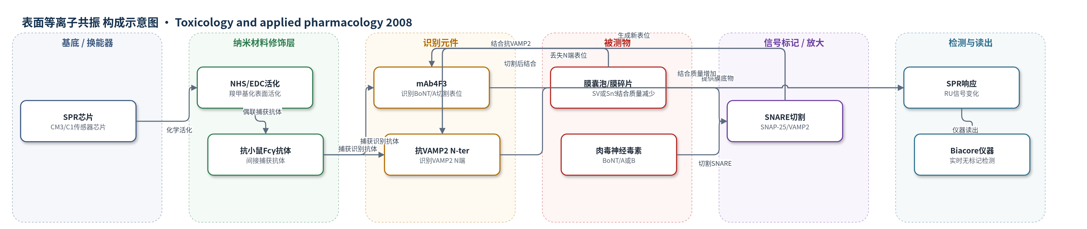
点击图片查看大图 · 依据论文自动绘制
传感器类型
表面等离子共振(SPR)生物传感器
检测对象
肉毒神经毒素A(Botulinum neurotoxin A, BoNT/A)、肉毒神经毒素B(Botulinum neurotoxin B, BoNT/B);样品基质:大鼠脑匀浆上清(SnH/SnS)、原代小脑颗粒神经元裂解液/细胞培养物
检测原理
SPR芯片经NHS/EDC活化并固定抗小鼠Fcγ抗体,再捕获抗VAMP2、抗SV2、抗SNAP-25或mAb4F3等识别抗体。样品中的突触囊泡或细胞膜碎片携带VAMP2/SNAP-25,被相应抗体捕获。BoNT/B轻链切割VAMP2 N端,使抗VAMP2 N-ter捕获下降;BoNT/A轻链切割SNAP-25 C端,产生新C端表位,使mAb4F3捕获上升。参考抗体信号用于归一化。膜蛋白大分子质量放大SPR响应,结合质量变化实时转换为共振单位(RU)信号,随毒素活性/浓度变化。
检测灵敏度
LOD: 4 pM SNAP-25(粗裂解液);绝对检测限: 2.7 pg SNAP-25(来自200 pg总蛋白);线性范围: 2–270 ng蛋白(CM3芯片);0.2–6 ng总蛋白(C1芯片);R^2 = 0.99;EC50: BoNT/B-Lc 1.1±0.1 pM(SPR)、1.3±0.2 pM(流式)、1.9±0.1 pM和1.1±0.1 pM(ELISA);BoNT/A-Lc 34±3 pM;完整神经元BoNT/A 571±192 fM。
效应效果
SPR与ELISA、流式细胞术对BoNT/B-Lc的EC50基本一致(SPR 1.1±0.1 pM,流式1.3±0.2 pM,ELISA 1.9±0.1和1.1±0.1 pM),且更快。非特异结合<10%;预孵育抗体阻断结合,100000×g离心消除信号,EDTA抑制BoNT/A-Lc,特异性高。膜结合稳定,可用辛基葡糖苷再生,同一芯片一周内可完成数百次注射。SPR灵敏度比ELISA高5–10倍,可检测<100 pg/ml膜抗原。96孔细胞实验中,200 fM BoNT/A使mAb4F3信号增加178±12%,100 pM BoNT/B使抗VAMP2信号下降43±7%。可替代小鼠致死试验,用于质量控制、抑制剂筛选和中和抗体检测。
传感器的构成
- SPR换能基底:CM3或C1传感器芯片,提供表面等离子共振检测表面
- 化学活化层:NHS/EDC混合液活化芯片羧甲基化表面,用于共价偶联抗体
- 捕获抗体层:兔多克隆抗小鼠Fcγ抗体(anti-mouse IgG),间接捕获单克隆抗体
- 识别元件:mAb4F3、抗VAMP2 N-ter、抗SV2、抗synaptophysin、抗SNAP-25 N-ter、抗syntaxin1、抗Na+/K+-ATPase抗体,分别识别SNARE表位或作参考
- 封闭剂:1 M ethanolamine,封闭剩余活化位点
- 样品底物:SnH(大鼠脑匀浆10000×g上清,含突触囊泡SV)、SnS(超声脑组织10000×g上清,含细胞膜SNAP-25)、神经元裂解液
- 信号标记物:无标记膜蛋白/囊泡,SPR直接检测结合质量变化
中文摘要
肉毒神经毒素A和B(BoNT/A、BoNT/B)通过裂解突触前SNARE蛋白SNAP-25和VAMP2阻断神经递质释放,既是危险病原体也是治疗/美容药物,因此需要体外活性检测替代小鼠试验。本研究利用表面等离子共振(SPR)检测抗体对膜囊泡的捕获,以评估毒素活性。以突触囊泡为底物,SPR、ELISA和流式细胞术测得的BoNT/B EC50相近,但SPR更快。超声破碎脑组织或神经元培养物可获得暴露胞内表位的细胞膜碎片,用于BoNT/A活性检测。SPR响应与抗原浓度成正比,可在粗裂解液中检测低至4 pM SNAP-25。使用特异性识别BoNT/A切割后SNAP-25表位的单克隆抗体,完整原代神经元中BoNT/A的EC50为0.5 pM。该SPR生物传感器方法可在96孔培养体系中监测BoNT/A和B活性,为毒理学检测提供替代动物实验的方法。
英文摘要
Botulinum neurotoxins A and B (BoNT/A and B) are neuromuscular blocking agents which inhibit neurotransmission by cleaving the intra-cellular presynaptic SNARE proteins SNAP-25 and VAMP2, localized respectively in plasma membrane and synaptic vesicles. These neurotoxins are both dangerous pathogens and powerful therapeutic agents with numerous clinical and cosmetic applications. Consequently there is a need for in vitro assays of their biological activity to screen for potential inhibitors and to replace the widely used in vivo mouse assay. Surface plasmon resonance (SPR) was used to measure membrane vesicle capture by antibodies against SNAP-25 and VAMP2. Substrate cleavage by BoNTs modified capture providing a method to assay toxin activity. Firstly using synaptic vesicles as a substrate, a comparison of the EC(50)s for BoNT/B obtained by SPR, ELISA or flow cytometry indicated similar sensitivity although SPR assays were more rapid. Sonication of brain or neuronal cultures generated plasma membrane fragments with accessible intra-cellular epitopes adapted to measurement of BoNT/A activity. SPR responses were proportional to antigen concentration permitting detection of as little as 4 pM SNAP-25 in crude lysates. BoNT/A activity was assayed using monoclonal antibodies that specifically recognize a SNAP-25 epitope generated by the proteolytic action of the toxin. Incubation of intact primary cultured neurons with BoNT/A yielded an EC(50) of 0.5 pM. The SPR biosensor method was sensitive enough to monitor BoNT/A and B activity in cells cultured in a 96-well format providing an alternative to experimental animals for toxicological assays.