组成图示
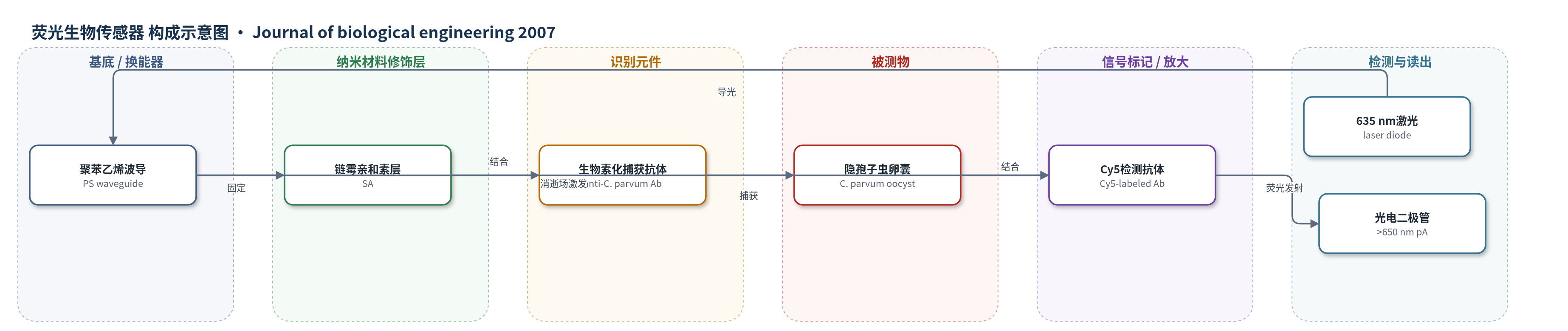
点击图片查看大图 · 依据论文自动绘制
传感器类型
荧光生物传感器
检测对象
微小隐孢子虫卵囊(Cryptosporidium parvum oocysts);样品基质:饮用水/水样(potable water/water samples,PBS模拟液、滤池反冲洗水)
检测原理
生物素化抗隐孢子虫卵囊抗体经链霉亲和素固定在聚苯乙烯波导表面,水样中的卵囊被捕获;Cy5标记检测抗体再结合卵囊表面抗原,形成免疫夹心。635 nm激光经波导产生消逝场,仅激发距表面约100–1000 nm内的Cy5,其>650 nm荧光部分耦合回波导,由光电二极管转换为pA信号。卵囊浓度越高,结合Cy5抗体越多,荧光/pA增量越大。煮沸可释放或暴露>300 kDa和105 kDa抗原,使更多Cy5靠近波导,从而提高灵敏度。
检测灵敏度
LOD: 89 ΔpA(多克隆抗体);110 ΔpA(单克隆抗体);检测浓度: 10^5 oocysts/ml(煮沸卵囊,多克隆抗体);10^6 oocysts/ml(未煮沸卵囊,多克隆抗体);灵敏度提高: 煮沸后多克隆抗体体系提高10倍
效应效果
多克隆抗体经ELISA、流式细胞术、免疫印迹和荧光显微镜验证。煮沸或冻融使多克隆ELISA信噪比至少提高5倍,胆汁或SDS约3倍,50、60、75°C处理10 min分别提高1.5、2.4、4倍。光纤传感器中,多克隆捕获/检测可检出煮沸卵囊10^5/mL和未煮沸10^6/mL;单克隆检测10^6/mL与PBS无显著差异。在滤池反冲洗水背景中,卵囊荧光明显高于碎片。RAPTOR试纸阴性时可重复使用最多20次。作者认为当前检出限高于EPA要求,需串联浓缩与热处理,可发展为饮用水在线自动监测。
传感器的构成
- 基底/换能器:聚苯乙烯(PS)光纤波导,传导635 nm激发光并产生消逝场,收集Cy5荧光
- 光阱:波导末端黑色涂料,吸收杂散光
- 亲和固定层:链霉亲和素(SA),固定生物素化捕获抗体
- 识别元件:生物素化抗隐孢子虫卵囊抗体(biotinylated anti-C. parvum oocyst antibody,多克隆/单克隆),捕获卵囊
- 信号标记物:Cy5标记检测抗体(Cy5-labeled anti-C. parvum oocyst antibody,多克隆/单克隆),结合卵囊表面抗原
- 封闭/检测缓冲液:酪蛋白(casein)和牛血清白蛋白(BSA)PBS缓冲液,降低非特异结合
- 读出模块:635 nm激光二极管与光电二极管,激发并检测>650 nm荧光,输出pA
中文摘要
微小隐孢子虫(Cryptosporidium parvum)是引起全球水源性胃肠疾病的重要肠道原虫,饮用水中卵囊检测对预防暴发至关重要。本研究评估抗隐孢子虫卵囊多克隆和单克隆抗体作为捕获与检测试剂,用于光纤生物传感器检测卵囊。抗体经酶联免疫吸附试验、流式细胞术、免疫印迹和荧光显微镜验证。当多克隆抗体同时用作捕获和检测试剂时,可检测10^5卵囊/mL;卵囊检测前煮沸可使多克隆抗体体系灵敏度提高10倍。免疫印迹和免疫荧光显示,单克隆和多克隆抗体均识别卵囊壁表面大于300 kDa的黏蛋白样抗原,多克隆抗体还识别煮沸样品中105 kDa小分子抗原。初步在线生物传感器系统设计表明,水样中的卵囊需先浓缩并热处理,才能被生物传感器有效检测。
英文摘要
An intestinal protozoan parasite, Cryptosporidium parvum, is a major cause of waterborne gastrointestinal disease worldwide. Detection of Cryptosporidium oocysts in potable water is a high priority for the water treatment industry to reduce potential outbreaks among the consumer populace. Anti-Cryptosporidium oocyst polyclonal and monoclonal antibodies were tested as capture and detection reagents for use in a fiber optic biosensor assay for the detection of Cryptosporidium oocysts. Antibodies were validated using enzyme-linked immunosorbent assays, flow cytometry, Western blotting and fluorescent microscopy. Oocysts could be detected at a concentration of 105 oocysts/ml when the polyclonal antibodies were used as the capture and detection reagents. When oocysts were boiled prior to detection, a ten-fold increase in sensitivity was achieved using the polyclonal antibody. Western blotting and immunofluorescence revealed that both the monoclonal and polyclonal antibodies recognize a large (>300 kDa) molecular weight mucin-like antigen present on the surface of the oocyst wall. The polyclonal antibody also reacted with a small (105 kDa) molecular weight antigen that was present in boiled samples of oocysts. Preliminary steps to design an in-line biosensor assay system have shown that oocysts would have to be concentrated from water samples and heat treated to allow detection by a biosensor assay.