组成图示
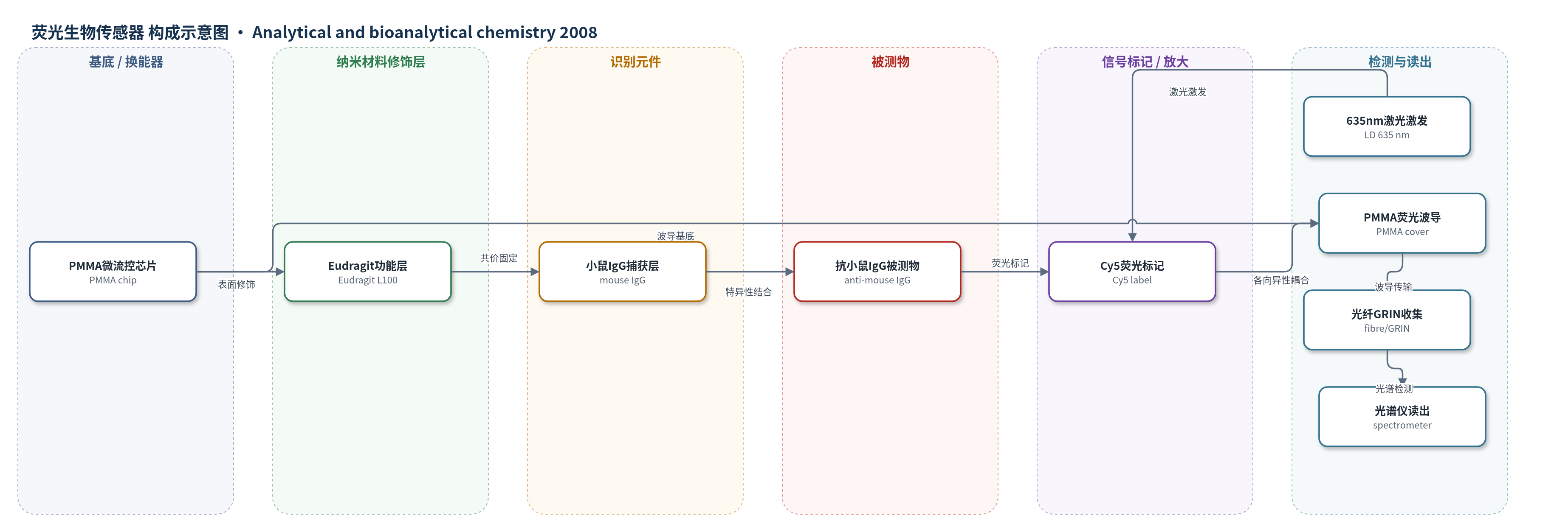
点击图片查看大图 · 依据论文自动绘制
传感器类型
荧光生物传感器
检测对象
Cy5 标记抗小鼠 IgG(anti-mouse IgG-Cy5);样品基质为 PBS 缓冲液(流动相)
检测原理
PMMA 上表面经 Eudragit L100 处理形成羧基,再用 EDC/NHS 活化,使小鼠 IgG 通过氨基-羧基共价键固定于盖板内壁。当含 Cy5 标记抗小鼠 IgG 的样品流过微通道时,抗小鼠 IgG 与固定 IgG 特异性结合,结合量随被测物浓度增加,使界面处 Cy5 荧光强度增加。635 nm 激光二极管激发 Cy5,发射荧光在 PMMA/液体界面具有各向异性,优先以约 60° 角度进入 PMMA 盖板并沿厚度方向波导传输,从端面经 GRIN 透镜和光纤收集,经 650 nm 长通滤光片后由光谱仪在 650–750 nm 积分。系统无酶催化放大,信号主要来自特异性免疫结合产生的荧光,背景包括 PMMA 自荧光和底部残留。
检测灵敏度
LOD: 0.05 μg mL−1;LOQ: 0.2 μg mL−1
效应效果
在 PBS 流动条件下,Eudragit L100 表面处理给出最高荧光响应,且非特异结合经 tris-HCl 封闭后可忽略,选择性较好。固定 IgG 浓度为 0.05 mg/mL、Eudragit L100 浓度为 0.01 mM 时响应最优。流动免疫孵育时间缩短至 15 min,PBS 洗涤 5 min,适合快速检测。加入柱面透镜、635 nm 带通滤光片并改用 Hamamatsu 光谱仪后,信噪比提高约一个数量级。校准点为 10 次光谱平均,标准差用于确定 LOD 和 LOQ。芯片为四通道结构,各通道光学隔离,潜在可用于多分析物检测。原文未报告实际生物样品加标回收率、长期稳定性、RSD 或与 ELISA/HPLC/qPCR 的定量对比。
传感器的构成
- 基底/换能器:聚甲基丙烯酸甲酯(PMMA)微流控芯片,由上下两片组成,下部含微通道与进出口,上部盖板作为荧光波导和传感层载体
- 表面功能化层:Eudragit L100(甲基丙烯酸/甲基丙烯酸酯共聚物)沉积于 PMMA 上表面,提供羧基(COOH)用于共价固定
- 活化交联层:EDC(1-乙基-3-[3-二甲基氨基丙基]碳二亚胺)和 NHS(N-羟基丁二酰亚胺)活化羧基,实现氨基与羧基共价连接
- 识别元件:小鼠 IgG(mouse IgG)共价固定于 PMMA 盖板内壁,作为捕获抗体
- 信号标记物:Cy5 标记抗小鼠 IgG(Cy5-labelled anti-mouse IgG),与固定 IgG 特异性结合并提供荧光
- 封闭剂:tris-HCl(0.2 M,pH 7.5)封闭未反应羧基,降低非特异结合;PBS 用于洗涤
- 流体/光学读出:PVC 管与蠕动泵输送 PBS,635 nm 激光二极管(LD)激发,GRIN 透镜与光纤收集,光谱仪检测
中文摘要
本文设计并开发了一种基于荧光各向异性的新型光学生物传感平台,用于读取光学生物芯片。该芯片由两片聚甲基丙烯酸甲酯(PMMA)加工而成,形成四条宽 500 μm、高 400 μm 的微通道;下部包含微通道及流体进出口,上部内表面固定传感生物层。光学信号来自生物层发射的荧光,该荧光在 PMMA/液体界面处呈各向异性耦合进入 PMMA 盖板,并沿芯片波导传输后被收集。作者通过微通道内直接 IgG/抗-IgG 相互作用验证其生物传感潜力:小鼠 IgG 共价固定于 PMMA 盖板内壁,Cy5 标记抗小鼠 IgG 作为特异性识别标记。研究比较了聚-L-乳酸(PLLA)、Eudragit L100 和 NaOH 等 PMMA 表面处理方法,以优化羧基分布并实现小鼠 IgG 共价固定;其中 Eudragit L100 处理效果最佳。所配置系统获得 0.05 μg/mL 的检出限和 0.2 μg/mL 的定量限。
英文摘要
A novel fluorescence-based optical platform for the interrogation of an optical biochip was designed and developed. The optical biochip was made of poly(methyl methacrylate) (PMMA) formed by two pieces of PMMA appropriately shaped in order to obtain four microchannels that are 500-microm wide and 400-microm high. The lower part includes the microchannels and the inlet and outlet for the fluidics, while the sensing biolayer was immobilized on the upper part. The optical signal comprised the fluorescence emitted by the biolayer, which was anisotropically coupled to the PMMA cover and suitably guided by the PMMA chip. The potentiality of the optical chip as a biosensor was investigated by means of a direct IgG/anti-IgG interaction carried out inside the flow channels. The mouse-IgG was covalently immobilized on the internal wall of the PMMA cover, and the Cy5-labelled anti-mouse IgG was used for the specific interaction. Several chemical treatments of the PMMA surface were investigated, poly(L: -lactic acid), Eudragit L100 and NaOH, in order to obtain the most effective distribution of carboxylic groups useful for the covalent immobilisation of the mouse-IgG. The treatment with Eudragit L100 was found to be the most successful. Limits of detection and quantification of 0.05 microg mL(-1) and 0.2 microg mL(-1), respectively, were obtained with the configuration described.