组成图示
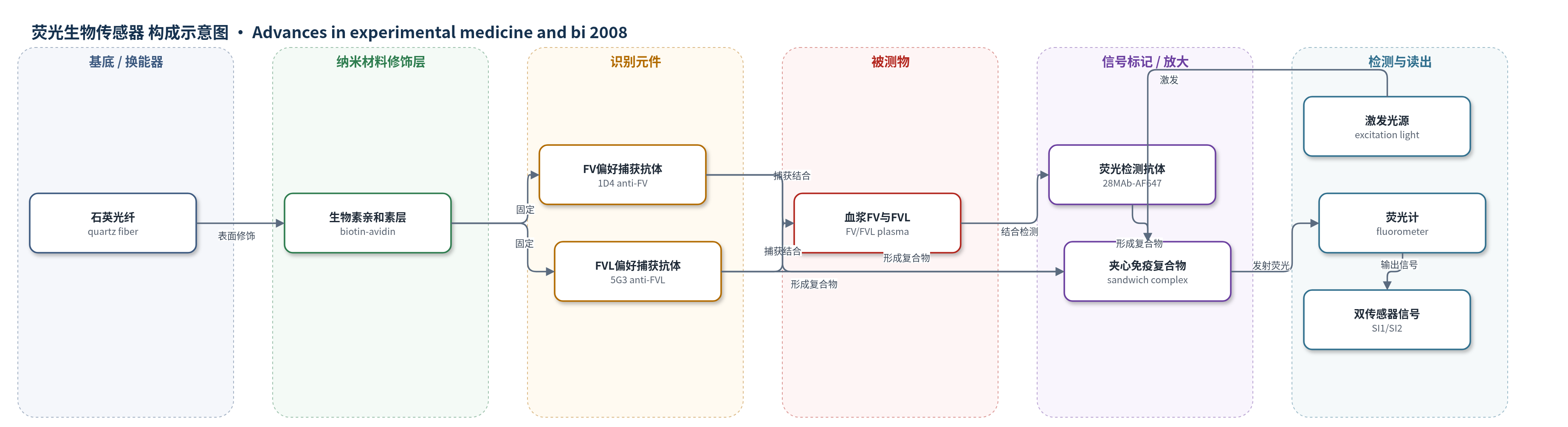
点击图片查看大图 · 依据论文自动绘制
传感器类型
荧光生物传感器
检测对象
凝血因子 V(Factor V, FV)、凝血因子 V Leiden 突变体(Factor V Leiden, FVL);样品基质:人血浆(blood plasma)
检测原理
传感器以石英光纤为基底,通过生物素-亲和素体系固定对 FV 或 FVL 具有偏好亲和力的捕获单克隆抗体 18MAb。血浆样品流经传感腔后,FV/FVL 被捕获抗体识别并结合;洗涤去除未结合蛋白后,加入 Alexa Fluor 647(AF647)标记的抗 FV 轻链检测抗体 28MAb,形成夹心免疫复合物。AF647 被激发光激发后发射荧光,荧光强度与结合在光纤表面的 FV/FVL 总量呈线性关系。由于两种捕获抗体均存在交叉反应,系统同时使用 FV 偏好型和 FVL 偏好型传感器,将两个荧光信号 SI1、SI2 与标准曲线斜率联立,解算出 FV 和 FVL 的浓度。该过程无核酸或酶催化放大,主要依靠双抗体夹心与荧光标记直接报告。
检测灵敏度
线性范围: 0–15 µg/ml;FV偏好传感器斜率: FV 9.3 pA/(µg/ml)、FVL 6.8 pA/(µg/ml);FVL偏好传感器斜率: FVL 8.6 pA/(µg/ml)、FV 5.4 pA/(µg/ml);混合样品斜率: FV偏好 8.9 pA/(µg/ml)、FVL偏好 8.4 pA/(µg/ml)
效应效果
两种传感器对 FV 和 FVL 均存在交叉反应,但混合样品中两种分子的信号贡献相互独立:FV 偏好传感器在固定 8 µg/ml FVL 时,FV 斜率为 8.9,为纯 FV 斜率 9.3 的 96%;FVL 偏好传感器在固定 8 µg/ml FV 时,FVL 斜率为 8.4,接近纯 FVL 斜率 8.6。示例血浆样品中 5 µg/ml FV 和 6 µg/ml FVL 被定量为 4.8±0.1 µg/ml 和 5.8±1.1 µg/ml,相对误差分别为 -4% 和 -3%,最大误差约 4%。完整检测约 8–10 分钟,明显快于传统 DNA 分析,且成本较低,可用于 FVL 诊断、严重程度评估及 FV 缺乏症筛查。
传感器的构成
- 基底/换能器:石英光纤(quartz fiber),传导激发光并收集 AF647 发射荧光。
- 表面连接层:生物素-亲和素(biotin-avidin)体系,将捕获抗体固定于光纤表面。
- 识别元件:捕获单克隆抗体 18MAb(1D4 或 5G3),分别构成 FV 偏好型和 FVL 偏好型传感器,识别血浆中 FV/FVL。
- 信号标记物:检测单克隆抗体 28MAb(抗 FV 轻链抗体)偶联 Alexa Fluor 647(AF647),形成夹心复合物并发出荧光。
- 反应腔:传感腔(sensing chamber),容纳血浆样品并实现孵育与洗涤。
- 读出装置:荧光计(fluorometer, Analyte 2000)与激发光,测量荧光强度并输出 SI1/SI2。
中文摘要
凝血因子 V Leiden(FVL)是凝血因子 V(FV)第 506 位精氨酸突变为谷氨酰胺导致的单氨基酸突变,是最常见的遗传性凝血障碍。目前 FVL 主要依靠 DNA 分析诊断,耗时长、成本高且需要专业人员。本文报道一种基于光纤表面夹心免疫反应的快速、准确、低成本生物传感系统,用于同时定量血浆中正常 FV 与突变 FVL,从而诊断 FVL 并评估疾病严重程度。为获得对 FV 或 FVL 具有偏好亲和力的单克隆抗体,作者将突变位点周围 20 肽分别偶联载体蛋白免疫小鼠,筛选杂交瘤细胞系。所得抗体虽存在交叉反应,但部分对 FV 亲和更高,部分对 FVL 亲和更高。据此构建 FV 偏好型和 FVL 偏好型两种光纤免疫荧光传感器,通过双传感器信号联立方程解算,可在约 8–10 分钟内完成检测,血浆样品中 FV 和 FVL 定量最大相对误差约 4%。该策略可推广至其他单点突变疾病的诊断。
英文摘要
Factor V Leiden (FVL) is an abnormality with a single amino acid mutation of Factor V (FV) and is the most common, hereditary blood coagulation disorder. FVL is currently diagnosed by DNA analysis, which takes a long assay time, high cost, and a specially trained person. We are developing a rapid, accurate, and cost-effective biosensing system to quantify both FV and FVL in blood plasma, to diagnose FVL and also to evaluate the seriousness of the disease status. This system is based on a sandwich immuno-reaction on an optical fiber. To produce the monoclonal antibody against only FV or only FVL without cross-reacting with the other molecule and with a higher probability, a 20 amino acid sequence (20-mer) of FV or FVL around the mutation region was injected into mice and then hybridoma cell lines specific to each 20-mer were selected. When these antibodies were tested with native FV or FVL molecules, they were found to be cross-reacting with the other molecules, but some with higher affinity to FV (FV preferred) and some to FVL (FVL preferred). Using these antibodies, two different sensors were developed: FV preferred and FVL preferred sensors. These two sensors allowed us to quantify FV and FVL in plasma with a maximum error of 4%. The plasma levels of both molecules provide us not only FVL diagnosis but also the level of the seriousness. The same principles may be used for developing diagnostic tools for other diseases with a single point mutation.