组成图示
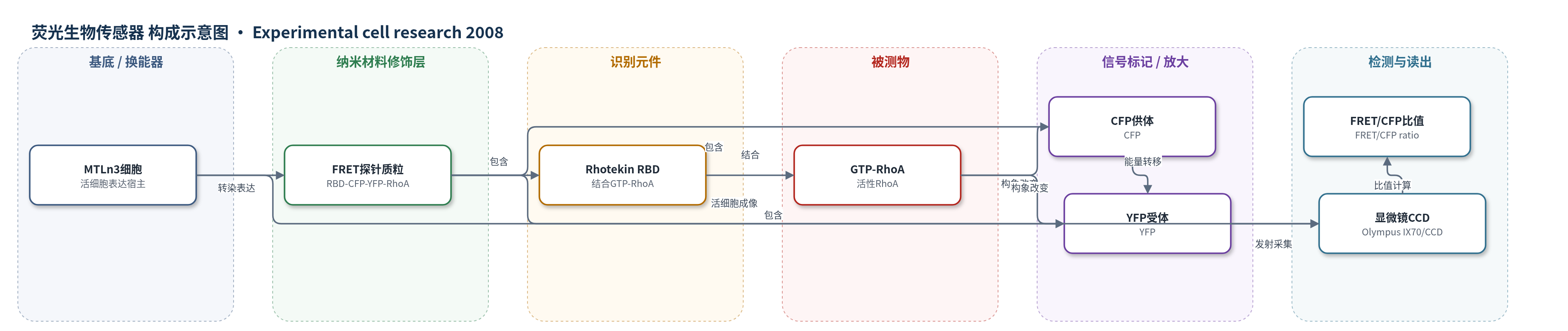
点击图片查看大图 · 依据论文自动绘制
传感器类型
荧光生物传感器
检测对象
活性RhoA(GTP-bound RhoA);样品基质:活细胞(MTLn3/MTC癌细胞胞质及前缘)和细胞裂解液(pull-down)
检测原理
该FRET生物传感器由Rhotekin RBD、CFP、17mer连接肽、YFP和全长RhoA串联组成。当RhoA结合GTP而激活时,RBD与GTP-RhoA结合,使CFP与YFP距离缩短,FRET效率升高。用430 nm激发CFP,分别采集CFP发射(470 nm)和YFP发射(535 nm),以FRET/CFP比值反映活性RhoA水平。EGF刺激MTLn3细胞后,前缘RhoA激活呈双相升高,比值成像可实时显示其时空分布。该法无需外源标记,信号随GTP-RhoA活性增加而增强,无化学放大步骤。
检测灵敏度
—
效应效果
该FRET探针经C3T微注射后信号受抑制,且表达不干扰EGF刺激的细胞骨架反应;分析限定中等表达(≤400 a.u./cell)以减少高表达伪影。时间序列成像显示EGF刺激后RhoA激活在1和3 min呈双相峰,并定位于前缘。抑制Rho或ROCK使迁移下降:C3T组总路径46.01±5.1 μm、净路径12.64±1.8 μm;Y27632组总路径36.45±1.2 μm、净路径7.25±0.7 μm,均低于对照。作者认为该传感器可实时解析RhoA在癌细胞前缘激活及其对Rac1/Cdc42突起调控的协调作用。
传感器的构成
- 表达载体:RhoA FRET生物传感器质粒,经Fugene6转染进入MTLn3/MTC细胞
- 识别元件:Rhotekin RBD,结合GTP-RhoA并触发构象变化
- 供体荧光蛋白:CFP,受430 nm激发,作为FRET供体
- 连接肽:17mer蛋白酶抗性无序连接肽,连接CFP与YFP并维持探针构象
- 受体荧光蛋白:YFP,接收CFP能量转移并在535 nm发射
- 报告靶标蛋白:全长RhoA,与RBD结合后改变CFP-YFP距离
- 光学读出:Olympus IX70显微镜与SensiCam QE CCD,采集CFP/FRET/YFP图像
- 数据分析:IP Lab软件计算FRET/CFP比值,定量RhoA活性
中文摘要
Rho GTP酶是调控细胞形态和肌动蛋白细胞骨架的关键分子开关。已有研究表明,Rac1和Cdc42分别调控迁移细胞前缘的板状伪足和丝状伪足形成,而RhoA主要介导细胞后部收缩;但近期报道发现迁移细胞前缘存在RhoA/ROCK激活区。本研究利用基于FRET的RhoA生物传感器,在EGF刺激的MTLn3癌细胞中实时监测RhoA活性,发现RhoA激活定位于细胞前缘,并呈双相动力学。抑制Rho或ROCK可增强膜突起,却显著降低细胞迁移能力,同时伴随细胞边缘Rac1明显激活。值得注意的是,对照MTLn3细胞中EGF刺激的突起不依赖Rac1而依赖Cdc42;ROCK抑制后则相反,突起依赖Rac1而不依赖Cdc42。结果表明,Rho和ROCK通过抑制前缘Rac1活性,协调Rac1与Cdc42对突起的调控,揭示Rho在癌细胞迁移信号中的新作用。
英文摘要
Rho GTPases are versatile regulators of cell shape that act on the actin cytoskeleton. Studies using Rho GTPase mutants have shown that, in some cells, Rac1 and Cdc42 regulate the formation of lamellipodia and filopodia, respectively at the leading edge, whereas RhoA mediates contraction at the rear of moving cells. However, recent reports have described a zone of RhoA/ROCK activation at the front of cells undergoing motility. In this study, we use a FRET-based RhoA biosensor to show that RhoA activation localizes to the leading edge of EGF-stimulated cells. Inhibition of Rho or ROCK enhanced protrusion, yet markedly inhibited cell motility; these changes correlated with a marked activation of Rac-1 at the cell edge. Surprisingly, whereas EGF-stimulated protrusion in control MTLn3 cells is Rac-independent and Cdc42-dependent, the opposite pattern is observed in MTLn3 cells after inhibition of ROCK. Thus, Rho and ROCK suppress Rac-1 activation at the leading edge, and inhibition of ROCK causes a switch between Cdc42 and Rac-1 as the dominant Rho GTPase driving protrusion in carcinoma cells. These data describe a novel role for Rho in coordinating signaling by Rac and Cdc42.