组成图示
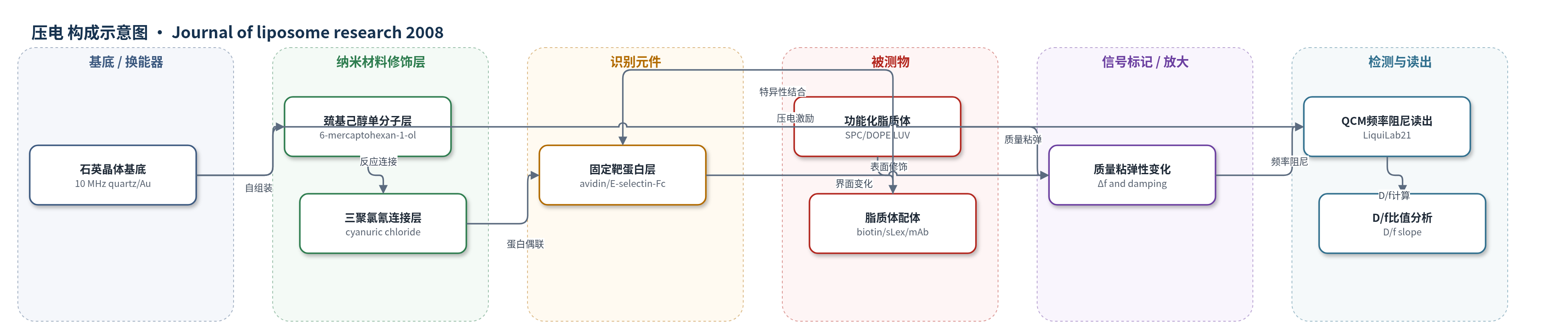
点击图片查看大图 · 依据论文自动绘制
传感器类型
压电(QCM)生物传感器
检测对象
非靶向 SPC 脂质体(SPC LUV)、生物素化脂质体(biotin-PE/biotin-PEG-PE LUV)、sLex 修饰脂质体(sLex-LUV)、抗 E-selectin 免疫脂质体(anti-E-selectin immunoliposomes)、DOPE-sLex 脂质体(DOPE-sLex LUV);样品基质:PBS 流动介质/模拟生理剪切流
检测原理
QCM 利用石英晶体压电振荡,表面质量增加使共振频率下降,按 Sauerbrey 方程与结合质量相关;表面粘弹性变化则引起阻尼频率改变。传感器金表面经 6-巯基己醇和三聚氯氰修饰后固定 avidin 或 E-selectin-Fc。流动 PBS 中,脂质体表面的 biotin、sLex 或抗 E-selectin 抗体与固定靶蛋白特异性结合,使脂质体吸附、变形、扁平化,甚至融合并释放内容物。结合质量增加导致 Δf 负向增大,脂质体由完整球体变为扁平/融合层时阻尼增加、D/f 斜率下降。高配体密度、高亲和 avidin/biotin 体系和 DOPE 促融合组分可放大变形/融合信号;PEG-PE 空间稳定则抑制变形。仪器实时读出 Δf 与阻尼,并通过 D/f 斜率判断脂质体状态。
检测灵敏度
原文未报告 LOD、线性范围、灵敏度斜率或相关系数。
效应效果
PBS剪切流下,非靶向SPC脂质体3 min内吸附,Δf约276±32 Hz,对应4.3 μg/cm²,D/f为2.83±0.4;PEG-PE降低非特异吸附,使Δf降至15±4 Hz、D/f升至3.51±0.7,疏水表面D/f为4.35±0.2,DOTAP脂质体Δf为938±23 Hz、D/f为2.29±0.6。生物素化脂质体20 mol%时Δf最大359±78 Hz,30 mol%时D/f降至0.98±0.01,提示铺展/融合;2%和5% PEG-PE使D/f升至3.72和4.02。sLex/DOPE脂质体2 h达最大结合,Δf约1200 Hz,D/f由3.36降至1.91,提示扁平化与内容物释放。数据为≥3次独立实验±SD,未报告RSD、回收率或LOD。
传感器的构成
- 基底/换能器:10 MHz 石英晶体(quartz crystal)及金表面(Au),压电振荡并响应表面质量与粘弹性变化
- 自组装单分子层:6-巯基己醇(6-mercaptohexan-1-ol)硫醇单分子层,提供反应端基并钝化表面
- 反应连接层:三聚氯氰(cyanuric chloride)反应性连接子,用于偶联靶蛋白
- 识别/靶蛋白层:亲和素(avidin)或重组人 E-selectin-Fc 嵌合蛋白,固定于金表面,特异性结合脂质体配体
- 流动池/介质:聚碳酸酯测量室(polycarbonate chamber)与 PBS 缓冲液,蠕动泵提供 270 μL/min 动态剪切流
- 分析物层:SPC/DOPE 脂质体(LUV),表面含 biotin-PE、biotin-PEG-PE、sLex-lipid 或经 N-glut-PE/EDC 偶联的抗 E-selectin 抗体,作为被测结合对象
中文摘要
本研究评估石英晶体微天平(QCM)作为生物传感器,在动态流动条件下实时研究脂质体与固定在传感器表面的靶蛋白之间的结合过程。QCM 的质量敏感频率变化可定量脂质体结合过程,同时阻尼分析可揭示脂质体在靶表面的变形或铺展程度。作者评估了一系列脂质体,其相互作用配体的种类和浓度不同,并与相应靶蛋白结合。结果表明,导向配体浓度升高会加速脂质体变形和扁平化,并触发融合过程。此外,比较生物素/亲和素与 E-selectin/配体相互作用发现,脂质体变形与靶分子结合亲和力相关。使用二油酰磷脂酰乙醇胺(DOPE)作为促融合膜组分可增强变形,而聚乙二醇磷脂酰乙醇胺(PEG-PE)的空间稳定作用则表现为较低变形。因此,QCM 在线检测脂质体靶结合,是控制和预测靶部位脂质体行为、提高治疗效力的优秀工具。
英文摘要
In this study we evaluated the quartz crystal microbalance (QCM) as a biosensor for a real-time investigation of liposomal binding, under dynamic flow conditions, onto target proteins immobilized at the sensor. The mass-sensitive frequency changes of quartz sensors allow for a quantification of the liposomal binding process. Furthermore, simultaneous damping analysis gives an insight into liposomal behavior, such as the degree of liposomal deformation or spreading at the target surface. In this study a series of liposomes was evaluated, differing in the kind and concentration of ligands interacting with appropriate target proteins. It became evident that an increase in homing device concentration accelerated deformation and flattening of liposomes, triggering a fusion process. Furthermore, liposomal deformation corresponded with the binding affinity of target molecules, comparing biotin/avidin with E-selectin/ligand interactions. Deformation could be emphasized using dioleoylphosphatidylethanolamine (DOPE) as a fusiogenic membrane component, while sterical stabilization by polyethylenglycol (PEG-PE) appeared in a low degree of deformation. Consequently, the online detection of liposomal target binding by QCM is an excellent facility to control and predict the liposomal behavior at the target site for increasing therapeutic potency.