组成图示
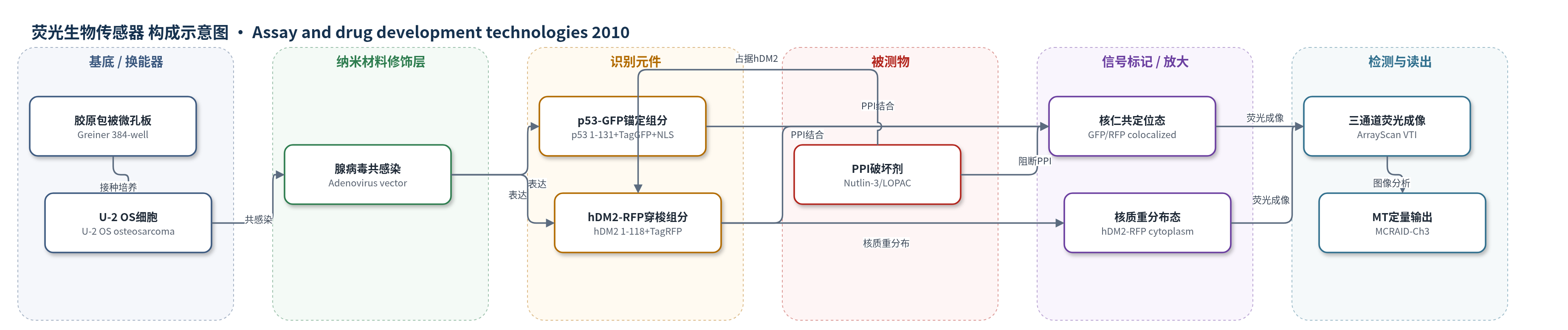
点击图片查看大图 · 依据论文自动绘制
传感器类型
荧光生物传感器
检测对象
p53-hDM2蛋白-蛋白相互作用破坏剂(p53-hDM2 protein–protein interaction disruptors,如Nutlin-3、mitoxantrone、ellipticine、camptothecin、topotecan);样品基质:U-2 OS细胞培养体系(McCoy's 5A培养基,≤0.5% DMSO,384孔板)
检测原理
重组腺病毒在U-2 OS细胞中表达p53 N端1–131融合TagGFP并含NLS,锚定于核仁;hDM2 N端1–118融合TagRFP并含NLS/NES,可在核质间穿梭。无破坏剂时,p53-GFP与hDM2-RFP的N端结构域结合,hDM2-RFP被锚定到核仁,GFP/RFP共定位,核内RFP平均强度高于胞质,MCRAID-Ch3较高。加入p53-hDM2 PPI破坏剂(如Nutlin-3)后,小分子占据hDM2 N端表面疏水口袋,阻断p53 N端Phe19/Trp23/Leu26与hDM2结合,hDM2-RFP从核仁释放并重新分布到胞质,核-胞质RFP强度差下降,MCRAID-Ch3随浓度降低。Hoechst 33342标记细胞核,ArrayScan VTI采集三通道图像,MT算法生成核/环掩膜并计算MCRAID-Ch3。该方法无酶放大,信号增强来自核仁锚定造成的强烈亚细胞重分布和荧光对比。
检测灵敏度
Nutlin-3 IC50: 0.607 – 0.382mM (n=5);R^2 = 0.97
效应效果
该PPIB HCS方法稳健:3天平均Z因子0.56±0.08、S:B 6.28±0.98;LOPAC四板Z′ 0.66、0.58、0.44、0.62,平均Z′ 0.57±0.11、S:B 8.7±0.7。DMSO>1%破坏图像分割,限定≤0.5%;Nutlin-3 5 min可见破坏,90 min为标准,信号稳定6 h且可逆。1280化合物LOPAC在50 mM下得9个(0.7%)潜在活性物,确认4个(mitoxantrone、ellipticine、camptothecin、topotecan)浓度依赖降低MCRAID-Ch3,但弱于Nutlin-3。作者另筛220,017化合物发现3个MBNA命中,IC50 30–50 mM,HCT116增殖抑制IC50约4 mM。
传感器的构成
- 检测基底:384孔胶原包被条码微孔板(Greiner collagen-coated barcoded microplate),承载细胞并适配ArrayScan VTI成像
- 细胞表达基质:U-2 OS骨肉瘤细胞,野生型p53、单拷贝hDM2,提供内源环境并作为成像背景
- 识别元件A:p53 N端1–131结构域融合TagGFP(p53-GFP),含NLS,锚定于核仁并提供hDM2结合位点
- 识别元件B:hDM2 N端1–118结构域融合TagRFP(hDM2-RFP),含NLS/NES,核质穿梭并在结合p53时锚定核仁
- 表达载体:重组腺病毒表达构建体(recombinant adenovirus constructs),分别携带p53-GFP与hDM2-RFP用于共感染表达
- 信号标记物:绿色荧光蛋白GFP(TagGFP)与红色荧光蛋白RFP(TagRFP),通过核仁共定位或核质重分布产生荧光信号
- 核标记/成像辅助:Hoechst 33342核酸染料,标记细胞核用于聚焦、核掩膜和分子转位(MT)图像分析
- 固定与读出:3.7%甲醛(formaldehyde)固定细胞,PBS洗涤后由ArrayScan VTI采集Hoechst/GFP/RFP三通道图像
中文摘要
本文报道一种新型成像型定位生物传感器高内容筛选(HCS)方法,用于鉴定p53-hDM2蛋白-蛋白相互作用(PPI)破坏剂。该生物传感器由嵌合蛋白组成,包含p53和hDM2的N端PPI结构域、核定位序列(NLS)、核输出序列(NES)及荧光报告蛋白。重组腺病毒共感染U-2 OS细胞后,表达p53 N端结构域融合绿色荧光蛋白(GFP)和hDM2 N端结构域融合红色荧光蛋白(RFP)。无破坏剂时,hDM2-RFP被锚定于核仁并与p53-GFP共定位;加入Nutlin-3后,其占据hDM2 N端表面疏水口袋,阻断与p53 N端结合,使hDM2-RFP重分布至胞质。作者以Nutlin-3验证该p53-hDM2 PPI生物传感器(PPIB)HCS方法,获得稳健、可重复且稳定的信号窗口。在1280化合物LOPAC筛选中,鉴定出4个能激活p53信号通路并产生明显区别于无活性化合物信号的化合物:米托蒽醌、椭圆精、喜树碱和拓扑替康。它们通过ATM-CHK2 DNA损伤应答通路激活p53,但破坏p53-hDM2 PPIB的效力远低于Nutlin-3,且表型不完全一致。
英文摘要
We present here the characterization and optimization of a novel imaging-based positional biosensor high-content screening (HCS) assay to identify disruptors of p53-hDM2 protein-protein interactions (PPIs). The chimeric proteins of the biosensor incorporated the N-terminal PPI domains of p53 and hDM2, protein targeting sequences (nuclear localization and nuclear export sequence), and fluorescent reporters, which when expressed in cells could be used to monitor p53-hDM2 PPIs through changes in the subcellular localization of the hDM2 component of the biosensor. Coinfection with the recombinant adenovirus biosensors was used to express the NH-terminal domains of p53 and hDM2, fused to green fluorescent protein and red fluorescent protein, respectively, in U-2 OS cells. We validated the p53-hDM2 PPI biosensor (PPIB) HCS assay with Nutlin-3, a compound that occupies the hydrophobic pocket on the surface of the N-terminus of hDM2 and blocks the binding interactions with the N-terminus of p53. Nutlin-3 disrupted the p53-hDM2 PPIB in a concentration-dependent manner and provided a robust, reproducible, and stable assay signal window that was compatible with HCS. The p53-hDM2 PPIB assay was readily implemented in HCS and we identified four (4) compounds in the 1,280-compound Library of Pharmacologically Active Compounds that activated the p53 signaling pathway and elicited biosensor signals that were clearly distinct from the responses of inactive compounds. Anthracycline (topoisomerase II inhibitors such as mitoxantrone and ellipticine) and camptothecin (topoisomerase I inhibitor) derivatives including topotecan induce DNA double strand breaks, which activate the p53 pathway through the ataxia telangiectasia mutated-checkpoint kinase 2 (ATM-CHK2) DNA damage response pathway. Although mitoxantrone, ellipticine, camptothecin, and topotecan all exhibited concentration-dependent disruption of the p53-hDM2 PPIB, they were much less potent than Nutlin-3. Further, their corresponding cellular images and quantitative HCS data did not completely match the Nutlin-3 phenotypic profile.