组成图示
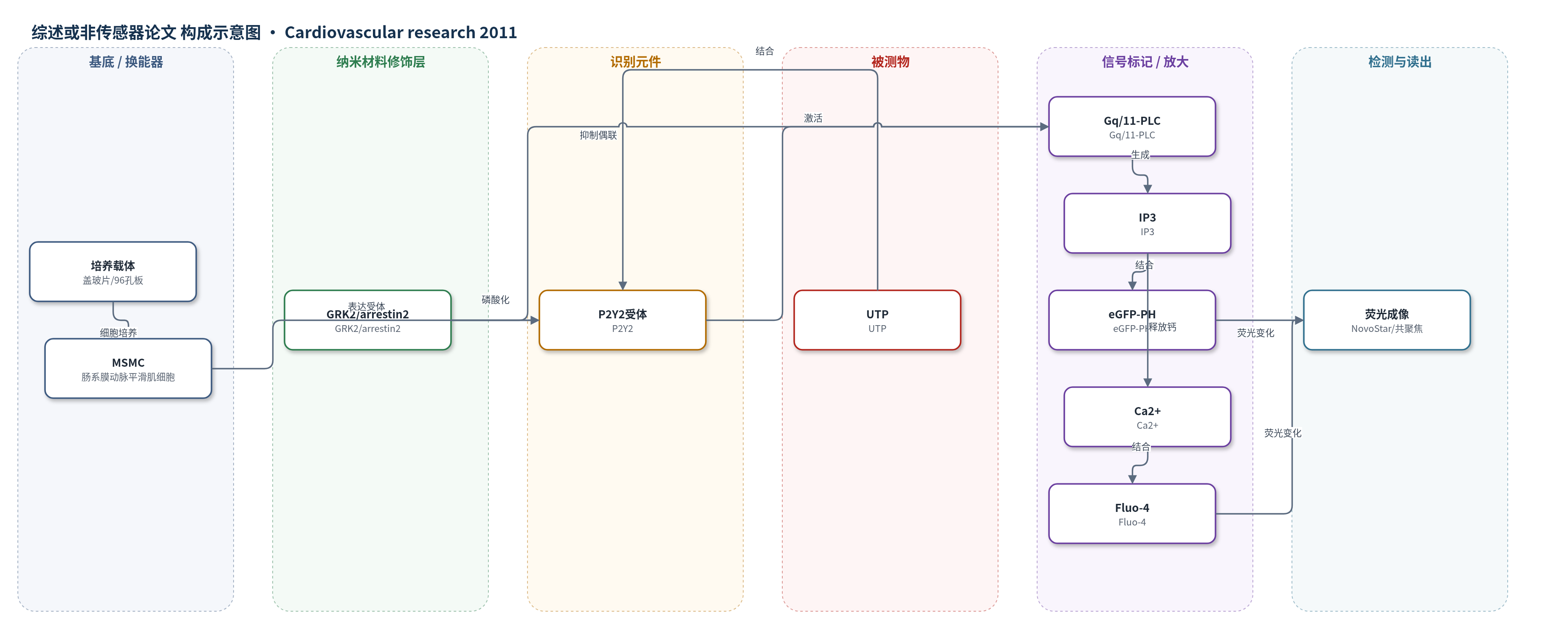
点击图片查看大图 · 依据论文自动绘制
传感器类型
综述或非传感器论文
检测对象
UTP(uridine 5′-triphosphate);样品基质:离体大鼠肠系膜动脉及原代肠系膜动脉平滑肌细胞(MSMCs)。
检测原理
UTP与MSMC膜上P2Y2受体结合后,激活Gq/11蛋白并刺激磷脂酶C(PLC),水解PIP2生成IP3和DAG。IP3一方面与eGFP-PH结合,引起其荧光强度变化,用于报告PLC活性;另一方面促使内质网释放Ca2+,使Fluo-4荧光增强,反映胞内钙信号。采用R1/Rmax/R2重复刺激方案时,最大浓度UTP先诱导受体脱敏:GRK2磷酸化P2Y2受体,arrestin2结合并阻断受体-G蛋白偶联,使第二次R2刺激的IP3和Ca2+信号显著降低。siRNA敲低GRK2或arrestin2可减弱该脱敏,而GRK3/5/6或arrestin3作用不明显。因此信号大小随UTP刺激状态和受体脱敏程度变化,而非简单线性浓度响应。
检测灵敏度
原文未报告 LOD、线性范围、灵敏度斜率或 R^2。
效应效果
研究在离体肠系膜动脉和原代MSMCs中显示UTP引起浓度依赖收缩,动脉EC50约28 μM(原文排版为28 mM),最大收缩在≥50 μM。Rmax后2 min,R2/R1收缩降低约80%,12–15 min恢复。细胞水平R2较R1的IP3信号降低84%,Ca2+信号降低64%。P2Y2 siRNA几乎完全消除UTP刺激的IP3/Ca2+信号,P2Y4 siRNA无影响,提示P2Y2为主。GRK2敲低≥75%或显性负突变使IP3 R2/R1由32±8%升至71±7%;arrestin2敲低>70%后脱敏明显减弱,arrestin3无显著作用。ETA受体脱敏则受arrestin3而非arrestin2调控,显示亚型选择性。作者认为GRK2/arrestin2是血管收缩信号调控的重要节点,可能用于高血压和血管疾病研究。
传感器的构成
- 基底/载体:盖玻片或96孔板(coverslips/96-well multiplates),用于细胞培养、成像与荧光检测。
- 识别元件:P2Y2嘌呤受体(P2Y2),位于MSMC膜上,识别UTP并激活下游PLC信号。
- 信号标记物:eGFP-PH(eGFP-PH,IP3生物传感器),结合IP3后引起荧光变化,报告PLC活性。
- 钙指示剂:Fluo-4-AM/Fluo-4,进入细胞后结合Ca2+,报告胞内钙浓度变化。
- 读出装置:NovoStar成像系统或共聚焦显微镜,读取F/F0荧光变化。
中文摘要
长期 P2Y 受体信号可导致血管收缩、高血压、血管平滑肌肥厚和增生。G 蛋白偶联受体信号受 G 蛋白偶联受体激酶(GRKs)和 arrestin 蛋白负向调控,防止持续或不当信号。本研究调查 GRKs 和 arrestins 是否调控成年 Wistar 大鼠肠系膜动脉平滑肌细胞(MSMCs)中 UTP 刺激的收缩信号。肠系膜动脉对 UTP 产生收缩;在最大 UTP 刺激前后分别给予 EC50 UTP(30 μM,原文排版为 30 mM,5 min),第二次反应较第一次降低,提示脱敏。利用转染 IP3 生物传感器 eGFP-PH 和/或加载 Ca2+ 敏感染料的分离 MSMCs 研究 UTP 诱导的 P2Y 受体 PLC 信号脱敏。类似方案显示 R2 反应较 R1 明显降低。通过转染显性负性 GRK 或靶向特定 GRK/arrestin 的 siRNA 探究其作用。GRK2 抑制(而非 GRK3、GRK5 或 GRK6)减弱 P2Y 受体脱敏;arrestin2 敲低减弱 UTP 刺激的 P2Y 受体脱敏,而 arrestin3 耗竭无此作用。P2Y2 受体特异性 siRNA 敲低几乎完全消除 UTP 刺激的 IP3/Ca2+ 信号,提示研究特异性表征该嘌呤受体亚型。这些数据强调 GRK2 和 arrestin2 是阻力动脉中 UTP 刺激的 P2Y2 受体反应性的重要调节因子,可能在血管疾病相关血管收缩信号通路中具有重要意义。
英文摘要
AIMS: prolonged P2Y-receptor signalling can cause vasoconstriction leading to hypertension, vascular smooth muscle hypertrophy, and hyperplasia. G protein-coupled receptor signalling is negatively regulated by G protein-coupled receptor kinases (GRKs) and arrestin proteins, preventing prolonged or inappropriate signalling. This study investigates whether GRKs and arrestins regulate uridine 5'-triphosphate (UTP)-stimulated contractile signalling in adult Wistar rat mesenteric arterial smooth muscle cells (MSMCs).
METHODS AND RESULTS: mesenteric arteries contracted in response to UTP challenge: When an EC(50) UTP concentration (30 µM, 5 min) was added 5 min before (R(1)) and after (R(2)) the addition of a maximal UTP concentration (R(max): 100 µM, 5 min), R(2) responses were decreased relative to R(1), indicating desensitization. UTP-induced P2Y-receptor desensitization of phospholipase C signalling was studied in isolated MSMCs transfected with an inositol 1,4,5-trisphosphate biosensor and/or loaded with Ca(2+)-sensitive dyes. A similar protocol (R(1)/R(2) = 10 µM; R(max) = 100 µM, applied for 30 s) revealed markedly reduced R(2) when compared with R(1) responses. MSMCs were transfected with dominant-negative GRKs or siRNAs targeting specific GRK/arrestins to probe their respective roles in P2Y-receptor desensitization. GRK2 inhibition, but not GRK3, GRK5, or GRK6, attenuated P2Y-receptor desensitization. siRNA-mediated knockdown of arrestin2 attenuated UTP-stimulated P2Y-receptor desensitization, whereas arrestin3 depletion did not. Specific siRNA knockdown of the P2Y(2)-receptor almost completely abolished UTP-stimulated IP(3)/Ca(2+) signalling, strongly suggesting that our study is specifically characterizing this purinoceptor subtype.
CONCLUSION: these new data highlight roles of GRK2 and arrestin2 as important regulators of UTP-stimulated P2Y(2)-receptor responsiveness in resistance arteries, emphasizing their potential importance in regulating vasoconstrictor signalling pathways implicated in vascular disease.