组成图示
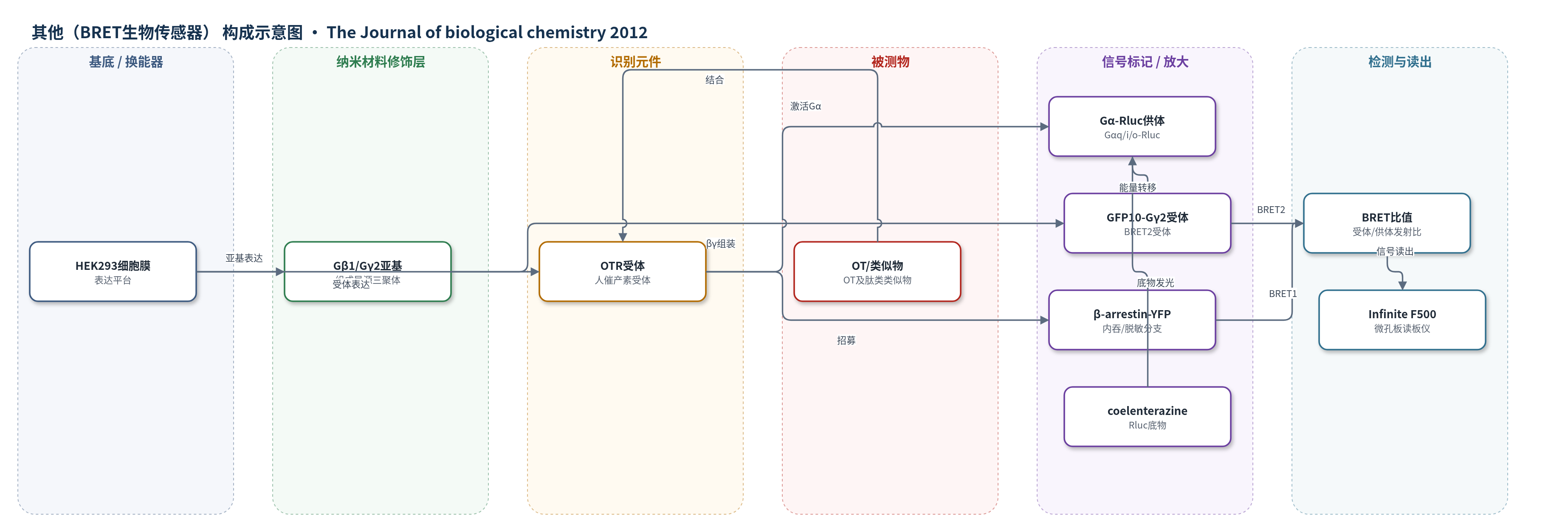
点击图片查看大图 · 依据论文自动绘制
传感器类型
其他(BRET生物传感器)
检测对象
催产素(oxytocin, OT)、催产素/血管加压素类似物(DNalOVT、atosiban、AVP、AVT 等);样品基质:HEK293 转染细胞悬液/细胞培养体系
检测原理
OT 或类似物结合 OTR 后,受体构象改变并招募/激活特定 Gαβγ。在 Gα-Rluc/GFP10-Gγ2 传感器中,Gα 与 Gβγ 分离使 Rluc 与 GFP10 距离增大,BRET 比值下降;在 OTR-Rluc/β-arrestin-YFP 传感器中,受体磷酸化后招募 β-arrestin,距离缩短,BRET 比值上升。加入 coelenterazine 400a 或 h 后,Rluc 生物发光通过非辐射能量转移激发荧光蛋白,读板仪分别检测供体与受体发射并计算比值。信号随配体浓度呈 EC50 依赖变化,Rluc8 可提高发光输出。
检测灵敏度
未报告 LOD、线性范围、灵敏度斜率或 R^2。报告 EC50: Gαq 2.16 ± 0.95 nM;Gαi1 62.63 ± 39.00 nM;Gαi2 32.27 ± 11.05 nM;Gαi3 11.50 ± 7.22 nM;GαoB 29.80 ± 19.32 nM;GαoA 91.80 ± 26.00 nM;DNalOVT-Gαi1 38.83 ± 16.0 nM;atosiban-Gαi3 2,800 ± 1,035 nM;β-arrestin1 229 ± 23.15 nM;β-arrestin2 41.15 ± 1.85 nM。
效应效果
BRET 法测得 OT 激活 Gαq 的 EC50 为 2.16 nM,与 IP1 法在子宫平滑肌细胞(1.4 nM)和 HEK293 细胞(1.7 nM)一致,验证传感器。OT 激活 Gq、Gi1、Gi2、Gi3、GoA、GoB,不激活 Gs;DNalOVT 仅激活 Gi1,atosiban 仅激活 Gi3,且不招募 β-arrestin 或诱导内化。100 nM 处理使 HEK293 增殖抑制 18.6±2、19±7、15.1±1.9%,DU145 抑制 27.9±1.3、27±0.2、24.1±0.7%;PTX 后为 1.8±2.1%、1.5±2%。未报告 RSD、稳定性或实际样品回收率。
传感器的构成
- 传感平台:HEK293 细胞,表达人 OTR 与 G 蛋白亚基,提供细胞内 BRET 检测环境
- 识别元件:OTR(OTR-Rluc、OTR-YFP 或 OTR-EGFP),结合 OT/AVP 类似物并传递激活信号
- G蛋白组装层:Gβ1 与 Gγ2(GFP10-Gγ2),组成异源三聚体并参与 BRET 报告
- 供体标记:Rluc/Rluc8 融合于 OTR C 端或插入 Gα 亚基(Gαq-97-Rluc、Gαi1-91-Rluc 等),产生生物发光
- 受体标记:GFP10-Gγ2 或 β-arrestin1/2-YFP,作为 BRET 能量受体,报告 Gα-Gγ 分离或 β-arrestin 招募
- 信号底物:coelenterazine 400a(CLz400)用于 BRET2,coelenterazine h 用于 BRET1,激发 Rluc 发光
- 读出装置:Infinite F500 微孔板读板仪,按 370–450/510–540 nm 或 370–480/520–570 nm 滤光片计算 BRET 比值
中文摘要
本研究利用生物发光共振能量转移(BRET)生物传感器筛选人催产素受体(OTR)的功能选择性配体。结果显示,催产素(OT)可促进 OTR 与 Gq 及所有 Gi/o 亚型直接结合并激活;其他在关键结构/功能残基上发生取代的肽类类似物均表现出对 G 蛋白亚型的偏向性激活。除 OT 外,没有配体能激活 GoA 或 GoB;除一种例外,所有激活 Gq 的肽也激活 Gi2 和 Gi3,但不激活 Gi1、GoA 或 GoB,显示强烈偏向。两种肽(DNalOVT 和 atosiban)分别仅激活 Gi1 或 Gi3,不招募 β-arrestin,也不诱导受体内化,首次明确区分 Gi/o 家族成员。两者均抑制细胞增殖,表明单一 Gi 亚型介导通路足以引发该生理反应。这些类似物为研究 Gi/o 成员在复杂生物学反应中的作用提供工具,并有助于开发具有新选择性和治疗特征的 OTR 靶向药物。
英文摘要
We used a bioluminescence resonance energy transfer biosensor to screen for functional selective ligands of the human oxytocin (OT) receptor. We demonstrated that OT promoted the direct engagement and activation of G(q) and all the G(i/o) subtypes at the OT receptor. Other peptidic analogues, chosen because of specific substitutions in key OT structural/functional residues, all showed biased activation of G protein subtypes. No ligand, except OT, activated G(oA) or G(oB), and, with only one exception, all of the peptides that activated G(q) also activated G(i2) and G(i3) but not G(i1), G(oA), or G(oB), indicating a strong bias toward these subunits. Two peptides (DNalOVT and atosiban) activated only G(i1) or G(i3), failed to recruit β-arrestins, and did not induce receptor internalization, providing the first clear examples of ligands differentiating individual G(i/o) family members. Both analogs inhibited cell proliferation, showing that a single G(i) subtype-mediated pathway is sufficient to prompt this physiological response. These analogs represent unique tools for examining the contribution of G(i/o) members in complex biological responses and open the way to the development of drugs with peculiar selectivity profiles. This is of particular relevance because OT has been shown to improve symptoms in neurodevelopmental and psychiatric disorders characterized by abnormal social behaviors, such as autism. Functional selective ligands, activating a specific G protein signaling pathway, may possess a higher efficacy and specificity on OT-based therapeutics.