组成图示
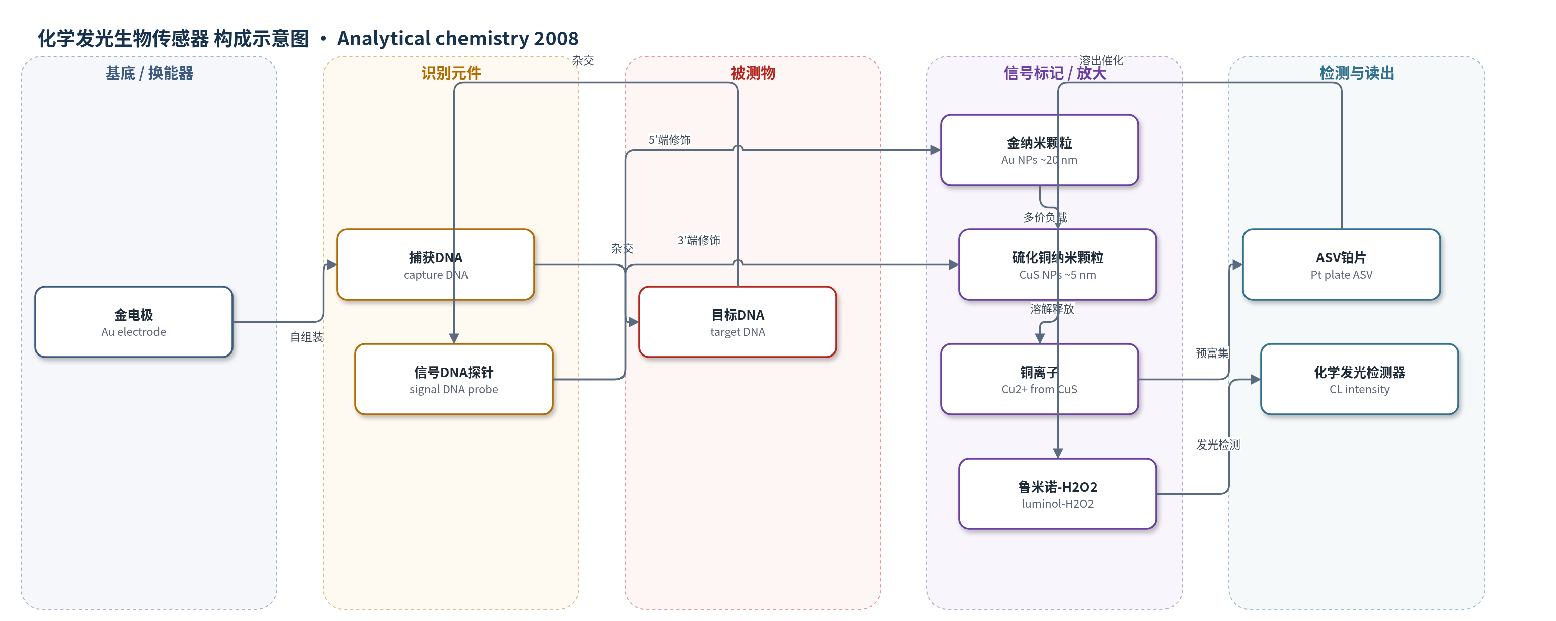
点击图片查看大图 · 依据论文自动绘制
传感器类型
化学发光生物传感器
检测对象
目标DNA(target DNA,序列特异性DNA);样品基质:PBS缓冲液/溶液(论文未涉及血清、尿液等实际生物基质)
检测原理
该传感器采用三明治杂交识别:巯基捕获DNA自组装于Au电极,目标DNA与捕获DNA杂交后,信号DNA探针再识别目标DNA另一端。信号探针5′端Au NPs可负载约10^1条探针,3′端CuS NPs作为Cu2+储库,实现多价信号放大。检测时,酸处理使CuS NPs溶解并释放Cu2+;Cu2+经ASV在Pt片上预富集后溶出,与luminol-H2O2反应。Cu2+催化鲁米诺氧化,产生与Cu2+浓度成正比的CL强度,从而间接反映目标DNA浓度。整体放大策略为Au NPs多价负载CuS标记探针与ASV预富集Cu2+。
检测灵敏度
LOD: 4.8 × 10^-15 M(3σ);线性范围: 2.0 × 10^-14–1.0 × 10^-13 M;灵敏度斜率: 14.6290(ICL对C,C以10^-14 M计);R = 0.9976;非线性范围: 2.0 × 10^-14–2.0 × 10^-12 M;R^2 = 0.9789。
效应效果
该传感器对互补目标DNA产生强CL信号,对双碱基错配DNA信号显著减弱,对非互补序列无响应,显示良好序列选择性。在4.0×10^-14 M目标DNA的11次重复测量中RSD为3.4%,重现性良好。LOD达4.8×10^-15 M,灵敏度较金纳米颗粒比色法提高约6个数量级,与Li等基于银纳米颗粒CL法(5 fM)相当,但步骤更简单、耗时更短。论文未报告实际生物样品加标回收率,作者认为其可用于高灵敏DNA杂交检测和免疫分析。
传感器的构成
- 基底电极:金电极(Au electrode),经抛光和电化学清洗,提供Au-S自组装界面并承载杂交结构。
- 捕获识别层:巯基功能化捕获DNA(capture DNA),通过Au-S键固定于Au电极,识别目标DNA一端。
- 封闭层:6-巯基-1-己醇(MCH),封闭裸露Au表面,降低后续杂交中的非特异吸附。
- 目标结合层:目标DNA(target DNA),与捕获DNA杂交形成双链,连接信号探针识别端。
- 信号识别层:信号DNA探针(signal DNA probe),识别目标DNA另一端,形成三明治型杂交体。
- 纳米放大标记:金纳米颗粒(Au NPs,约20 nm),修饰于信号DNA 5′端,可负载约10^1条探针以放大CuS数量。
- 发光前体标记:硫化铜纳米颗粒(CuS NPs,约5 nm),修饰于信号DNA 3′端,酸溶后释放Cu2+催化发光。
- 化学发光试剂:鲁米诺(luminol)与过氧化氢(H2O2),作为CL底物,在Cu2+催化下产生发光信号。
- 预富集电极:铂片(Pt plate),用于阳极溶出伏安法(ASV)预富集和溶出Cu2+,增强CL信号。
中文摘要
本文报道了一种基于纳米颗粒信号放大的新型高灵敏流注射化学发光(FI-CL)序列特异性DNA检测方法。该“三明治型”DNA生物传感器以巯基功能化捕获DNA自组装于金电极表面,并与目标DNA一端杂交;目标DNA另一端由信号DNA探针识别,该探针分别在3′端和5′端标记硫化铜纳米颗粒(CuS NPs)和金纳米颗粒(Au NPs)。杂交事件通过溶解杂合体中CuS释放的Cu2+催化鲁米诺-过氧化氢(luminol-H2O2)体系的化学发光强度进行监测。Au NPs可负载数百条带CuS NPs的信号DNA探针,从而显著提高灵敏度与选择性;金电极上Au NPs、信号DNA探针和CuS NPs的比例约为1/10^1/10^3。阳极溶出伏安法(ASV)对Cu2+进行预富集进一步提升了性能。在最优条件下,CL强度随目标DNA浓度在2.0×10^-14–2.0×10^-12 M范围内增加,检出限达4.8×10^-15 M,并对双碱基错配DNA表现出优异选择性。
英文摘要
A novel and sensitive flow injection chemiluminescence assay for sequence-specific DNA detection based on signal amplification with nanoparticles (NPs) is reported in the present work. The "sandwich-type" DNA biosensor was fabricated with the thiol-functionalized capture DNA first immobilized on an Au electrode and hybridized with one end of target DNA, the other end of which was recognized with a signal DNA probe labeled with CuS NPs and Au NPs on the 3'- and 5'-terminus, respectively. The hybridization events were monitored by the CL intensity of luminol-H2O2-Cu(2+) after the cupric ions were dissolved from the hybrids. We demonstrated that the incorporation of Au NPs in this sensor design significantly enhanced the sensitivity and the selectivity because a single Au NP can be loaded with hundreds of signal DNA probe strands, which were modified with CuS NPs. The ratios of Au NPs, signal DNA probes, and CuS NPs modified on the gold electrode were approximately 1/101/103. A preconcentration process of cupric ions performed by anodic stripping voltammetry technology further increased the sensor performance. As a result of these two combined effects, this DNA sensor could detect as low as femtomolar target DNA and exhibited excellent selectivity against two-base mismatched DNA. Under the optimum conditions, the CL intensity was increased with the increase of the concentration of target DNA in the range of 2.0 x 10(-14)-2.0 x 10(-12) M. A detection limit of 4.8 x 10(-15) M target DNA was achieved.